==============================================
⬇️✨⬇️🎉⬇️🔥⬇️📚⬇️
Click the link to purchase on Amazon 🎉📚
==============================================
🎥 Check Out All Videos at Once! 📺
👉 Visit Visualizing MSK Blog to explore a wide range of videos! 🩻
https://visualizingmsk.blogspot.com/?view=magazine
📚 You can also find them on MSK MRI Blog and Naver Blog! 📖
https://www.instagram.com/msk_mri/
Click now to stay updated with the latest content! 🔍✨
==============================================
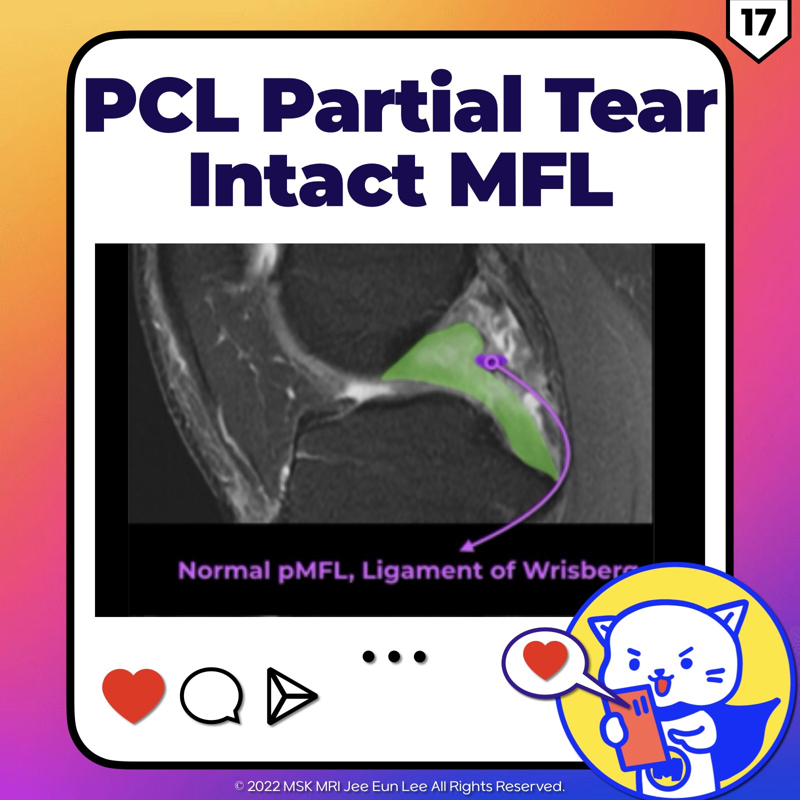
🔴Partial Tear of PCL
1️⃣Frequency and Diagnosis Challenges:
- Partial tears are more common in the PCL than in the ACL.
- An acutely torn PCL can appear as a continuous structure in 62% to 75% of cases, making it difficult to differentiate between complete and partial tears.
- Reference: Magn Reson Imaging Clin N Am. 2022 May;30(2):261-275.
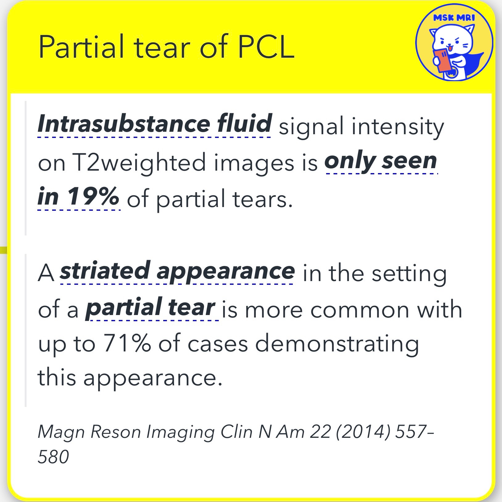
2️⃣Imaging Characteristics:
- Intrasubstance fluid signal intensity on T2-weighted images is observed in only 19% of partial tears.
- A striated appearance is more common in partial tears, with up to 71% of cases demonstrating this appearance.
- Reference: Magn Reson Imaging Clin N Am 22 (2014) 557–580.
https://visualizingmsk.blogspot.com/?view=flipcard
"Visualizing MSK Radiology: A Practical Guide to Radiology Mastery"
© 2022 MSK MRI Jee Eun Lee All Rights Reserved.
#VisualizingMSK #PCLinjuries #KneeMRI #PCLtear #MFL #Wrisberg #Humphrey
You should not distribute or commercially exploit the content.
You should not transmit or store it on any other website or electronic retrieval system.


'✅ Knee MRI Mastery > Chap 2.ACL and PCL' 카테고리의 다른 글
| (Fig 2-E.20) PCL Deficient Knee with Delayed Imaging (0) | 2024.03.18 |
|---|---|
| (Fig 2-E.19) PCL Bony Avulsion At Tibial Insertion (0) | 2024.03.17 |
| (Fig 2-E.16) Bone Contusions In PCL Hyperextension Injury (2) | 2024.03.16 |
| (Fig 2-E.14) Common MRI Findings of PCL Tears (0) | 2024.03.16 |
| (Fig 2-E.13) Transverse Tear of PCL And Impingement (0) | 2024.03.16 |