Click the link to purchase on Amazon 🎉📚
==============================================
🎥 Check Out All Videos at Once! 📺
👉 Visit Visualizing MSK Blog to explore a wide range of videos! 🩻
https://visualizingmsk.blogspot.com/?view=magazine
📚 You can also find them on MSK MRI Blog and Naver Blog! 📖
https://www.instagram.com/msk_mri/
Click now to stay updated with the latest content! 🔍✨
==============================================
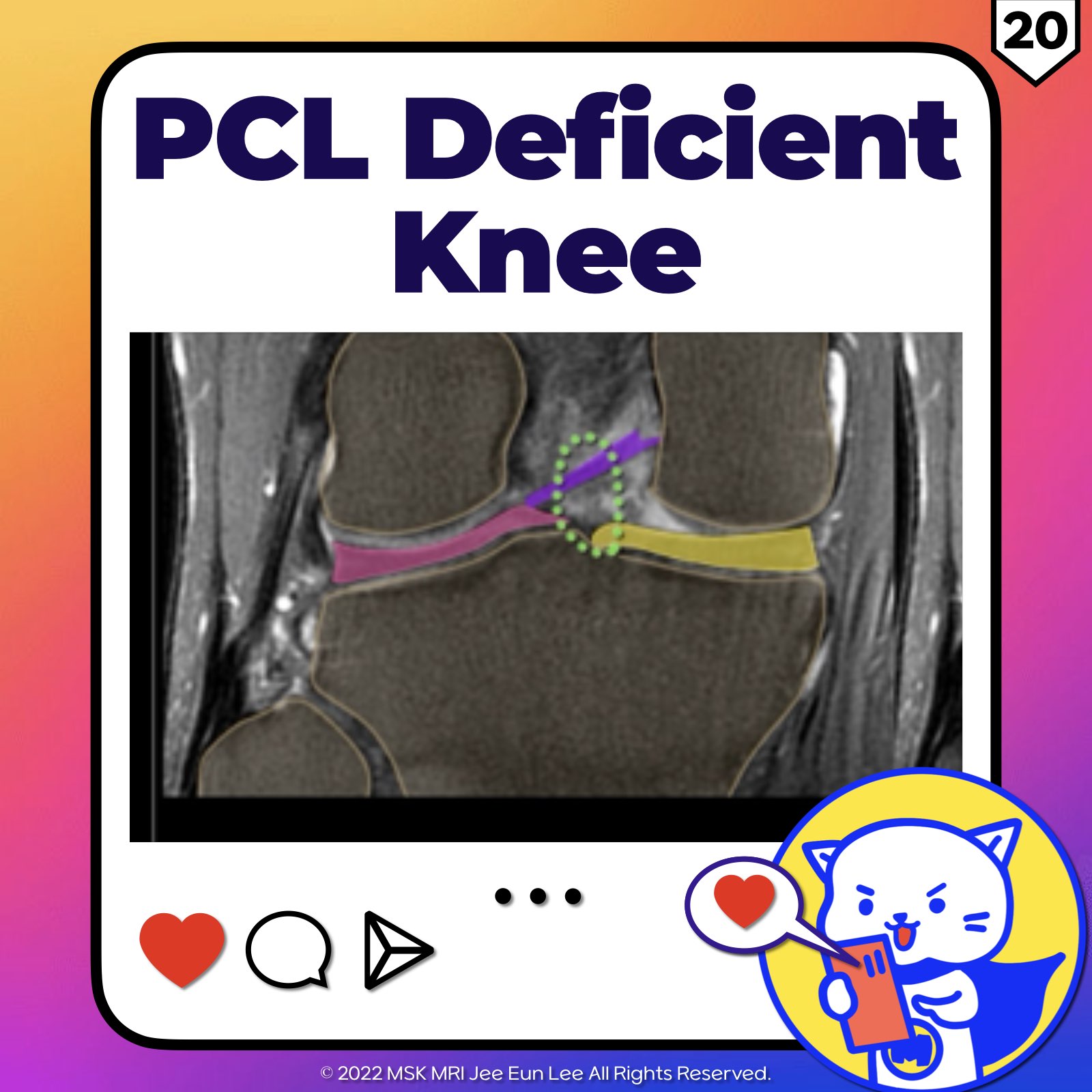
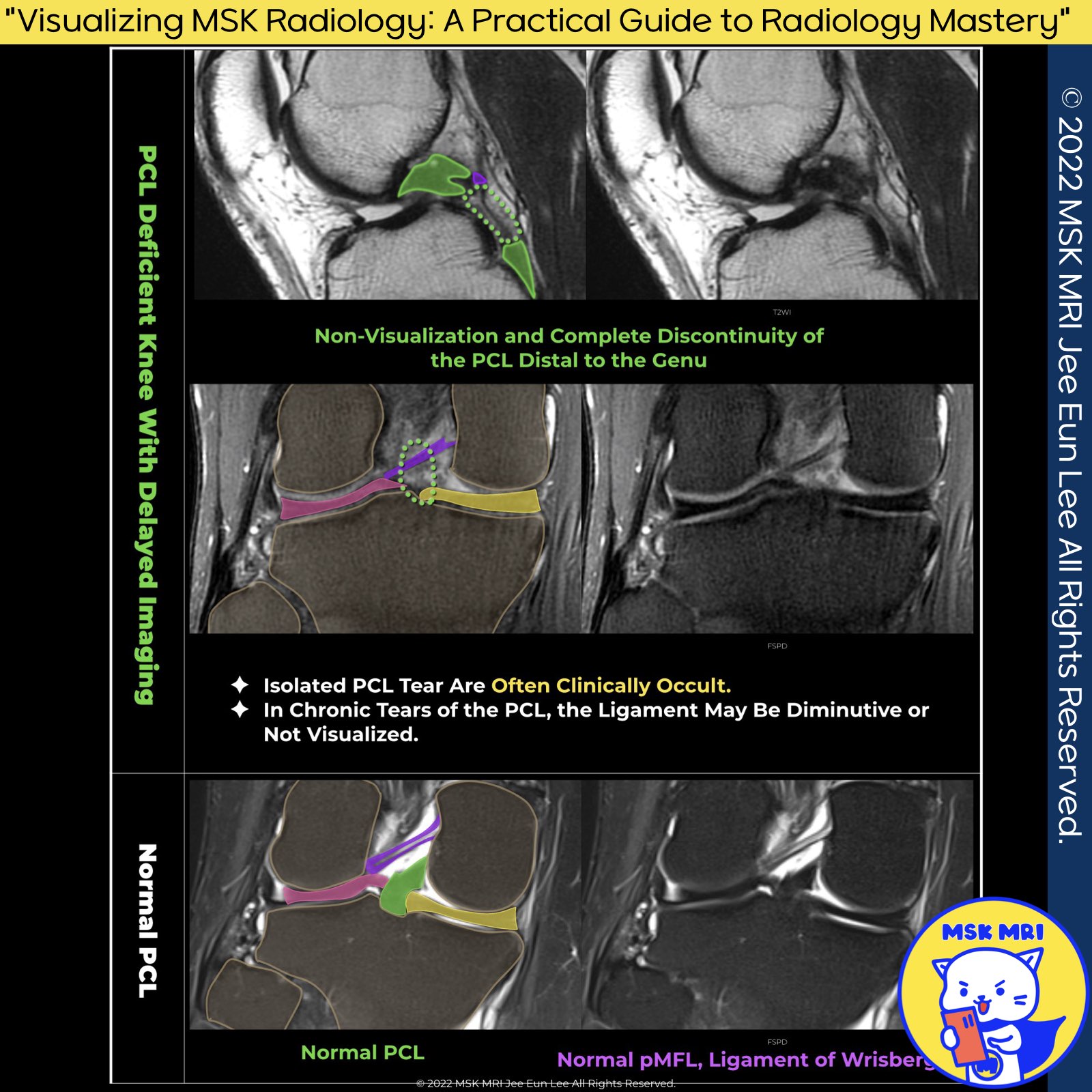
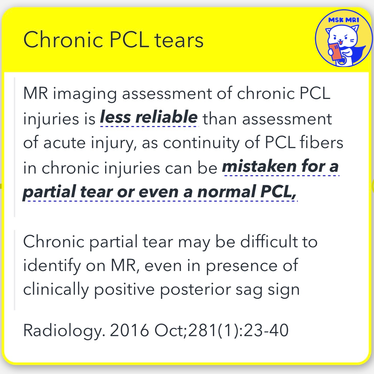
1️⃣ Chronic PCL Tears: MR Imaging Insights
✅MR imaging for chronic PCL injuries is less reliable, often confusing ligament continuity with partial or no tears. Identifying chronic partial tears is difficult, even with clinical indicators.
Source: Radiology, Oct 2016; 281(1):23-40.
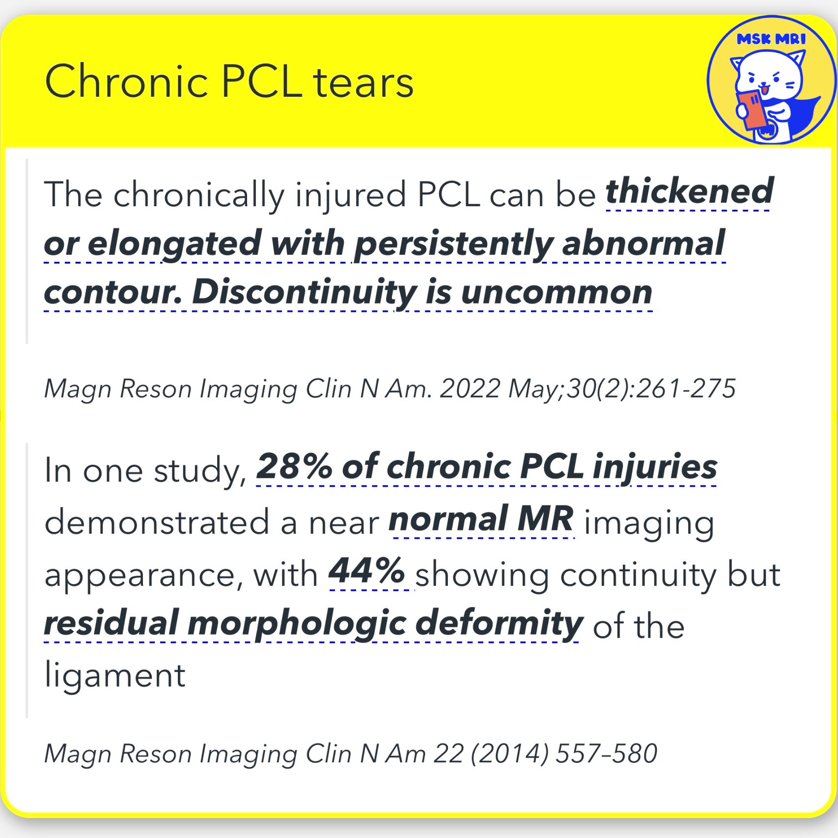
✅ Chronic PCL damage may show ligament thickening or elongation, with abnormal shape but seldom discontinuity.
Source: Magn Reson Imaging Clin N Am, May 2022; 30(2):261-275.
✅In studies, 28% of chronic PCL injuries appeared nearly normal on MRI, while 44% showed ligament continuity with morphologic abnormalities.
Source: Magn Reson Imaging Clin N Am 2014; 22:557–580.
2️⃣ Chronic PCL Injuries and Associated Chondral Lesions:
✅ About 80% of chronic cases exhibit chondral lesions on the medial femoral condyle and patellofemoral areas.
✅ Patients tend to lock their knees in extension to prevent posterior tibial translation, potentially leading to patellofemoral chondral changes over time.
Source: Radiology, Oct 2016; 281(1):23-40.
https://visualizingmsk.blogspot.com/?view=flipcard
"Visualizing MSK Radiology: A Practical Guide to Radiology Mastery"
© 2022 MSK MRI Jee Eun Lee All Rights Reserved.
#VisualizingMSK #PCLinjuries #KneeMRI #PCLtear #ChronicPCLinjury #PCL
You should not distribute or commercially exploit the content.
You should not transmit or store it on any other website or electronic retrieval system.


'✅ Knee MRI Mastery > Chap 2.ACL and PCL' 카테고리의 다른 글
| (Fig 2-E.22) Posterior Tibial Translation (0) | 2024.04.28 |
|---|---|
| (Fig 2-E.21) Radiographic Assessment of Posterior Knee Laxity (0) | 2024.04.27 |
| (Fig 2-E.19) PCL Bony Avulsion At Tibial Insertion (0) | 2024.03.17 |
| (Fig 2-E.17) Partial Tears of PCL and Intact MFL (0) | 2024.03.16 |
| (Fig 2-E.16) Bone Contusions In PCL Hyperextension Injury (2) | 2024.03.16 |