==============================================
⬇️✨⬇️🎉⬇️🔥⬇️📚⬇️
Click the link to purchase on Amazon 🎉📚
==============================================
🎥 Check Out All Videos at Once! 📺
👉 Visit Visualizing MSK Blog to explore a wide range of videos! 🩻
https://visualizingmsk.blogspot.com/?view=magazine
📚 You can also find them on MSK MRI Blog and Naver Blog! 📖
https://www.instagram.com/msk_mri/
Click now to stay updated with the latest content! 🔍✨
==============================================
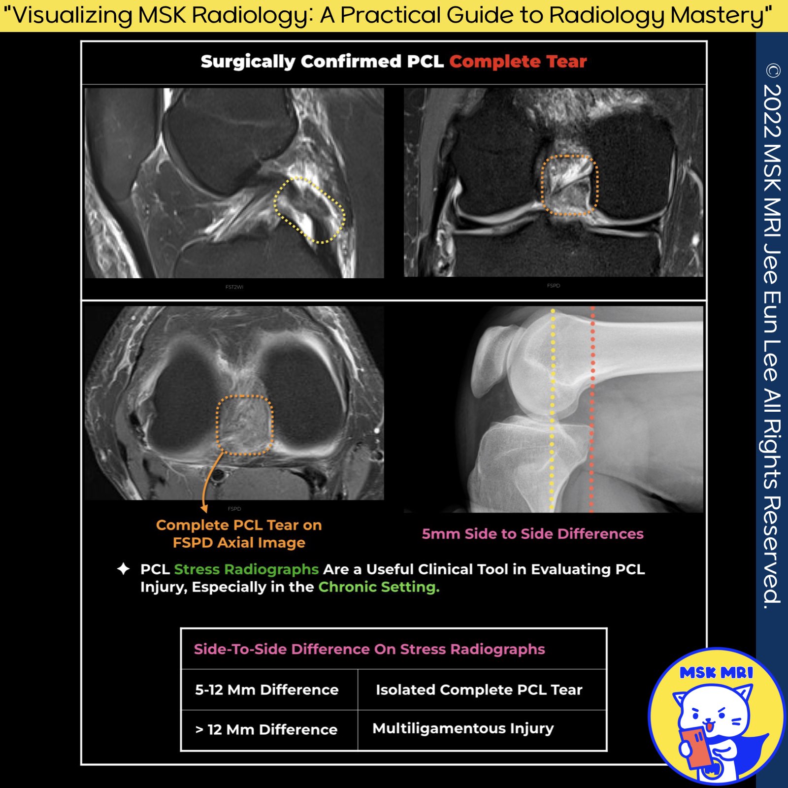
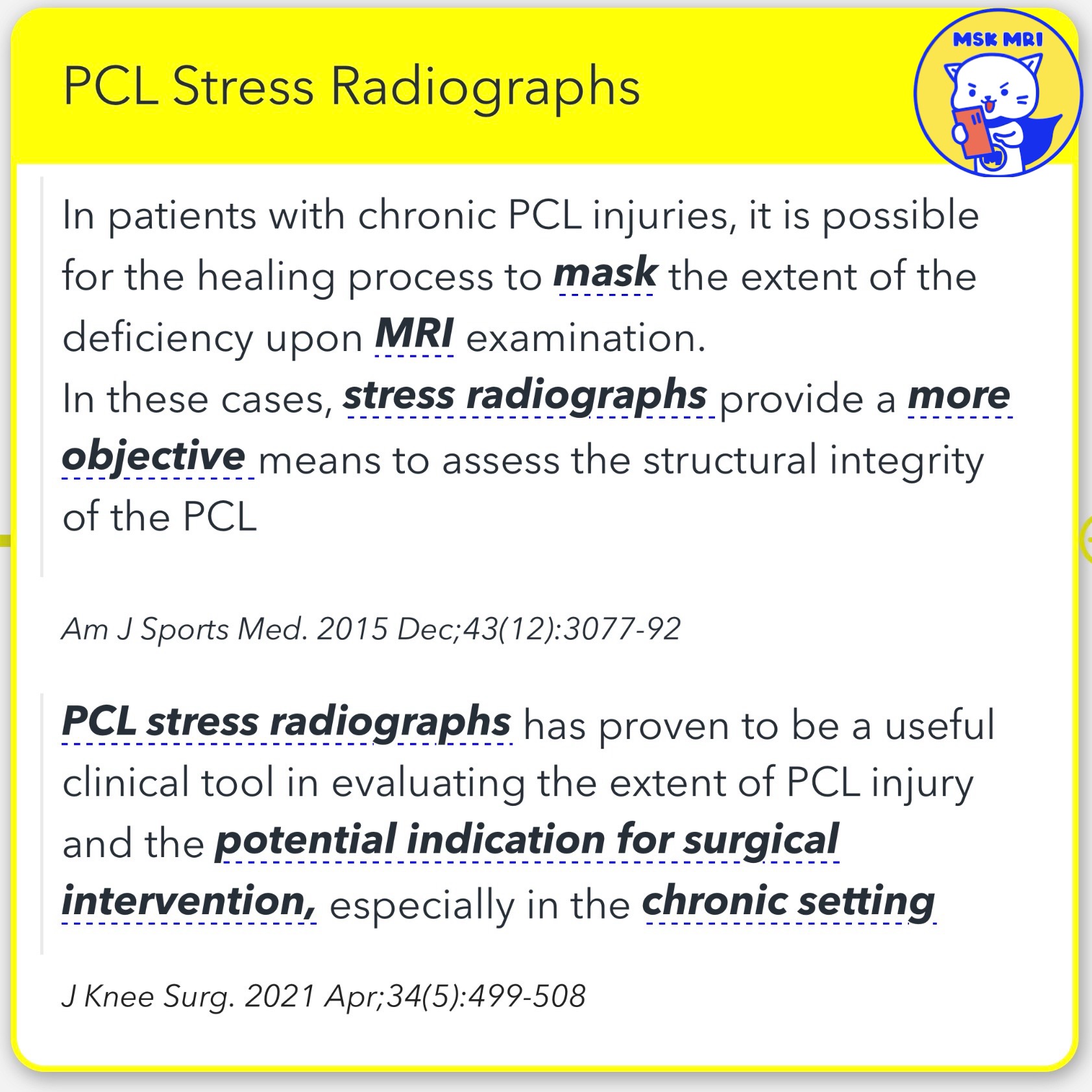
1️⃣ PCL Stress Radiographs in Chronic Injuries:
- Due to the healing process, MRI examinations in patients with chronic PCL injuries might not fully reveal the extent of the deficiency.
- Stress radiographs offer a more objective method to assess the PCL's structural integrity.
- American Journal of Sports Medicine, December 2015; 43(12): 3077-92.
2️⃣Clinical Utility of PCL Stress Radiographs:
- These radiographs are useful clinical tools for evaluating the extent of PCL injury and determining the need for surgical intervention, especially in chronic conditions.
- Journal of Knee Surgery, April 2021; 34(5): 499-508.
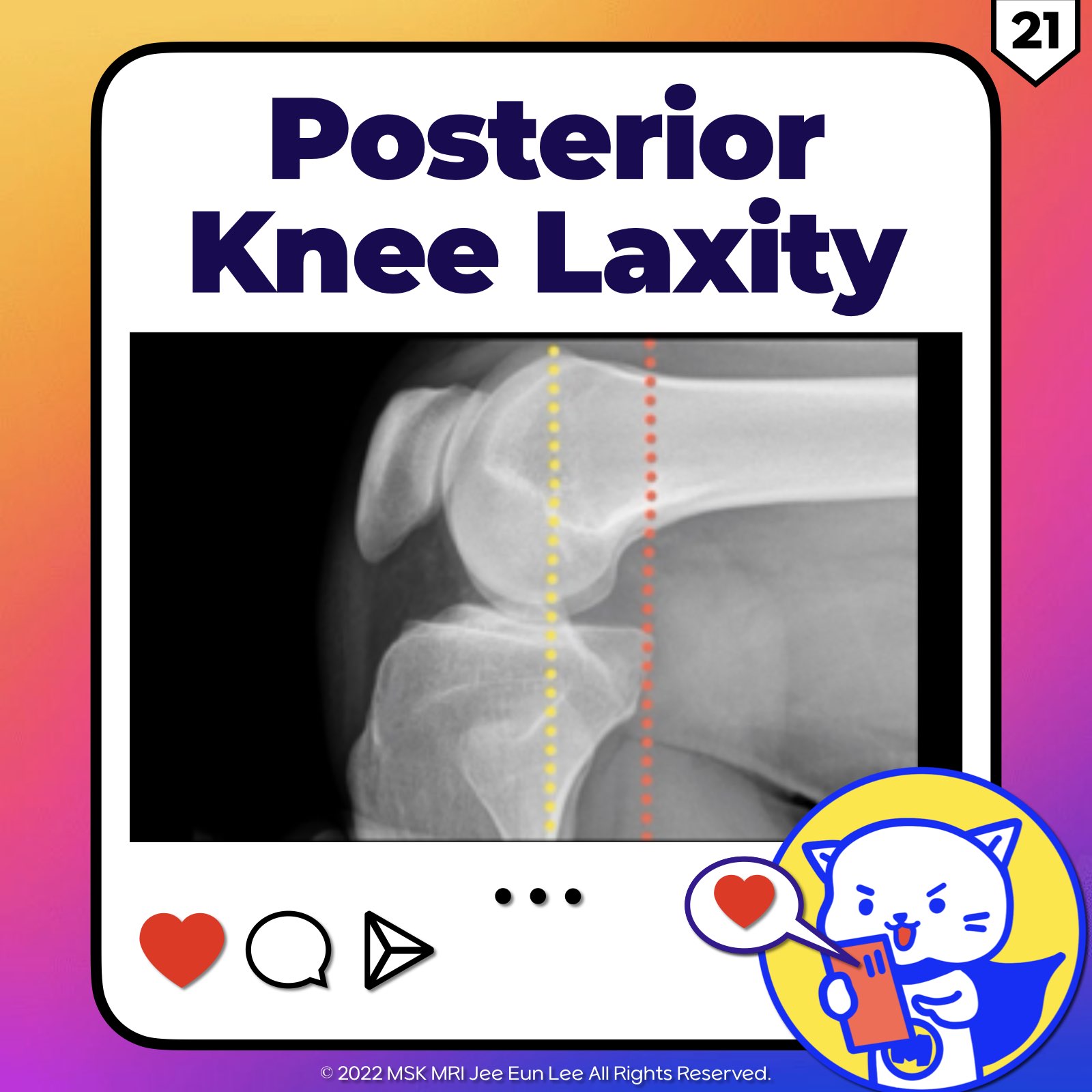
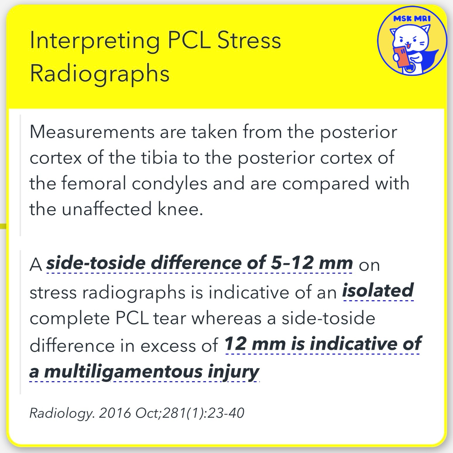
3️⃣Interpreting PCL Stress Radiographs:
- Method: Measurements are taken from the tibia's posterior cortex to the femoral condyles' posterior cortex, compared with the unaffected knee.
- Findings:
- Radiology, October 2016; 281(1): 23-40.
4️⃣ Enhanced Detection of PCL Injury with Proton Density Images
- Limitation of T2-Weighted Images: Sole reliance on these images may overlook subtle pathological changes in the PCL, resulting in decreased sensitivity.
- The advantage of Proton Density Images is that they offer enhanced visibility of abnormal intermediate or fluid intrasubstance signals, making them superior for detecting subtle PCL changes.
Magn Reson Imaging Clin N Am. 2022 May;30(2):261-275
AJR Am J Roentgenol. 2008 Oct;191(4):1031
"Visualizing MSK Radiology: A Practical Guide to Radiology Mastery"
© 2022 MSK MRI Jee Eun Lee All Rights Reserved.
No unauthorized reproduction, redistribution, or use for AI training.
#pcl #pclinjury #kneemri #mskmri #visualizingmsk


'✅ Knee MRI Mastery > Chap 2.ACL and PCL' 카테고리의 다른 글
| (Fig 2-E.23) Chronic Complete Tear of the PCL - Part 1 (2) | 2024.04.28 |
|---|---|
| (Fig 2-E.22) Posterior Tibial Translation (0) | 2024.04.28 |
| (Fig 2-E.20) PCL Deficient Knee with Delayed Imaging (0) | 2024.03.18 |
| (Fig 2-E.19) PCL Bony Avulsion At Tibial Insertion (0) | 2024.03.17 |
| (Fig 2-E.17) Partial Tears of PCL and Intact MFL (0) | 2024.03.16 |