Click the link to purchase on Amazon 🎉📚
==============================================
🎥 Check Out All Videos at Once! 📺
👉 Visit Visualizing MSK Blog to explore a wide range of videos! 🩻
https://visualizingmsk.blogspot.com/?view=magazine
📚 You can also find them on MSK MRI Blog and Naver Blog! 📖
https://www.instagram.com/msk_mri/
Click now to stay updated with the latest content! 🔍✨
==============================================
Chronic posterior cruciate ligament (PCL) injuries can present with various MRI appearances, posing challenges in accurately diagnosing and assessing functional integrity.
📌Key points regarding chronic PCL complete tears:
1️⃣Morphological Changes:
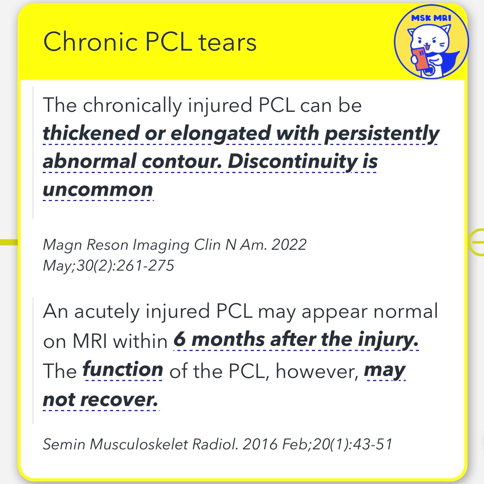
- The chronically injured PCL may appear thickened, elongated, or have a persistently abnormal contour on MRI.
- Discontinuity of the ligament is uncommon in chronic cases.
2️⃣Diminutive or Non-visualization:
- In some chronic tears, the PCL may appear diminutive or not visualized on MRI.
3️⃣Near-normal Appearance:
- A significant proportion (28% in one study) of chronic PCL injuries may demonstrate a near-normal MRI appearance, despite functional deficiency.
4️⃣Residual Morphologic Deformity:
- In many cases (44% in one study), the PCL may appear continuous but with residual morphologic deformity, indicating a previous injury.
✅Limitations of MRI:
- MRI has limitations in detecting chronically injured and functionally deficient PCL, as the ligament's appearance may not accurately reflect its functional status.
Magn Reson Imaging Clin N Am. 2022 May;30(2):261-275
Radiology. 2016 Oct;281(1):23-40.
Semin Musculoskelet Radiol. 2016 Feb;20(1):43-51
Radiology. 2016 Oct;281(1):23-40.
"Visualizing MSK Radiology: A Practical Guide to Radiology Mastery"
© 2022 MSK MRI Jee Eun Lee All Rights Reserved.
No unauthorized reproduction, redistribution, or use for AI training.


'✅ Knee MRI Mastery > Chap 2.ACL and PCL' 카테고리의 다른 글
| (Fig 2-E.25) Multiligamentous Injuries Associated with PCL Injuries (0) | 2024.04.28 |
|---|---|
| (Fig 2-E.24) Chronic Complete Tear of the PCL - Part 2 (0) | 2024.04.28 |
| (Fig 2-E.22) Posterior Tibial Translation (0) | 2024.04.28 |
| (Fig 2-E.21) Radiographic Assessment of Posterior Knee Laxity (0) | 2024.04.27 |
| (Fig 2-E.20) PCL Deficient Knee with Delayed Imaging (0) | 2024.03.18 |