==============================================
⬇️✨⬇️🎉⬇️🔥⬇️📚⬇️
Click the link to purchase on Amazon 🎉📚
==============================================
🎥 Check Out All Videos at Once! 📺
👉 Visit Visualizing MSK Blog to explore a wide range of videos! 🩻
https://visualizingmsk.blogspot.com/?view=magazine
📚 You can also find them on MSK MRI Blog and Naver Blog! 📖
https://www.instagram.com/msk_mri/
Click now to stay updated with the latest content! 🔍✨
==============================================
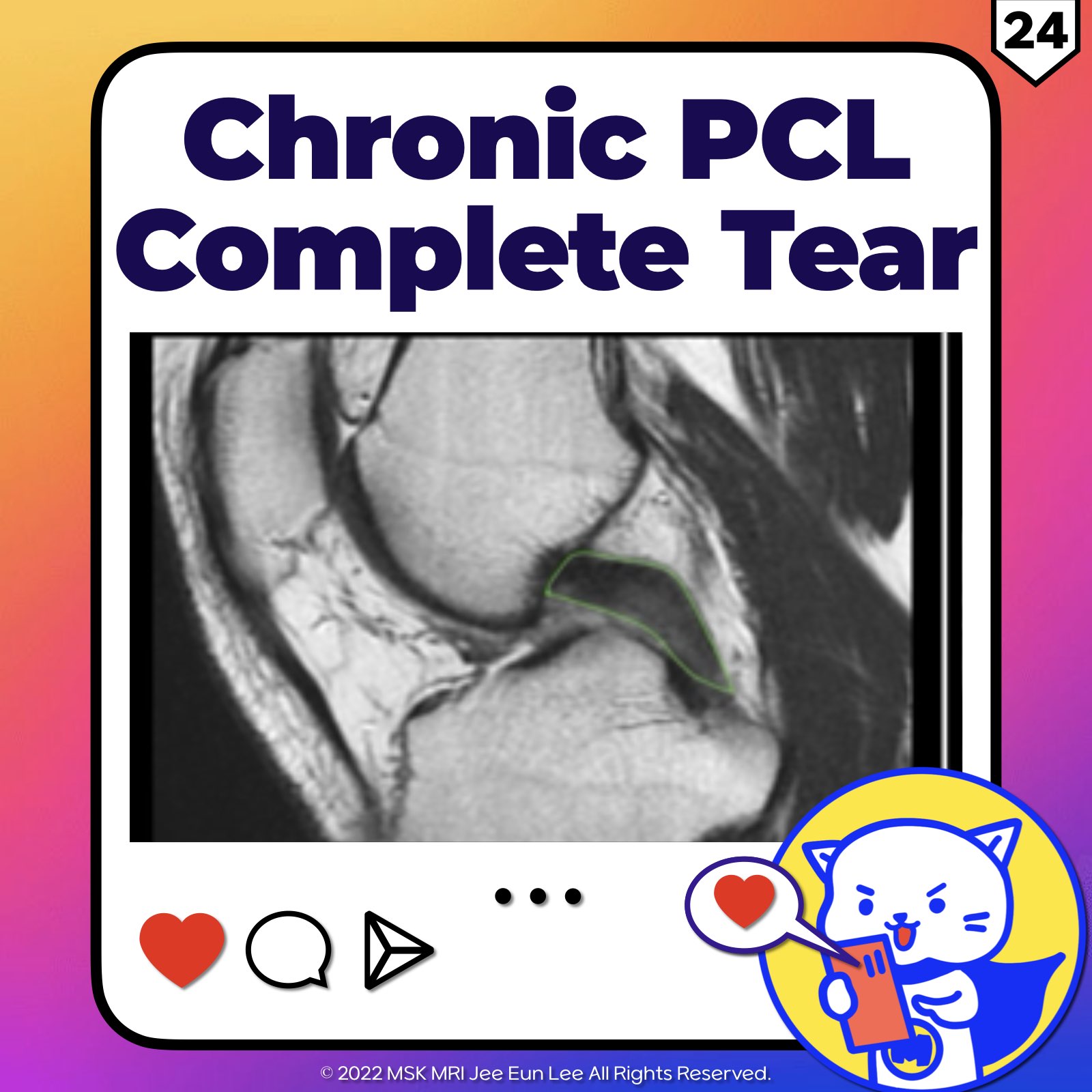
📌 Diagnosing Chronic PCL Tears
1️⃣ : Understanding PCL Appearance on MRI
- Chronic tears of the Posterior Cruciate Ligament (PCL) often present subtle signs that can be challenging to detect on MRI.
- Studies indicate that nearly 28% of chronic PCL injuries might appear almost normal, while another 44% may demonstrate continuity but with noticeable residual morphologic deformities.
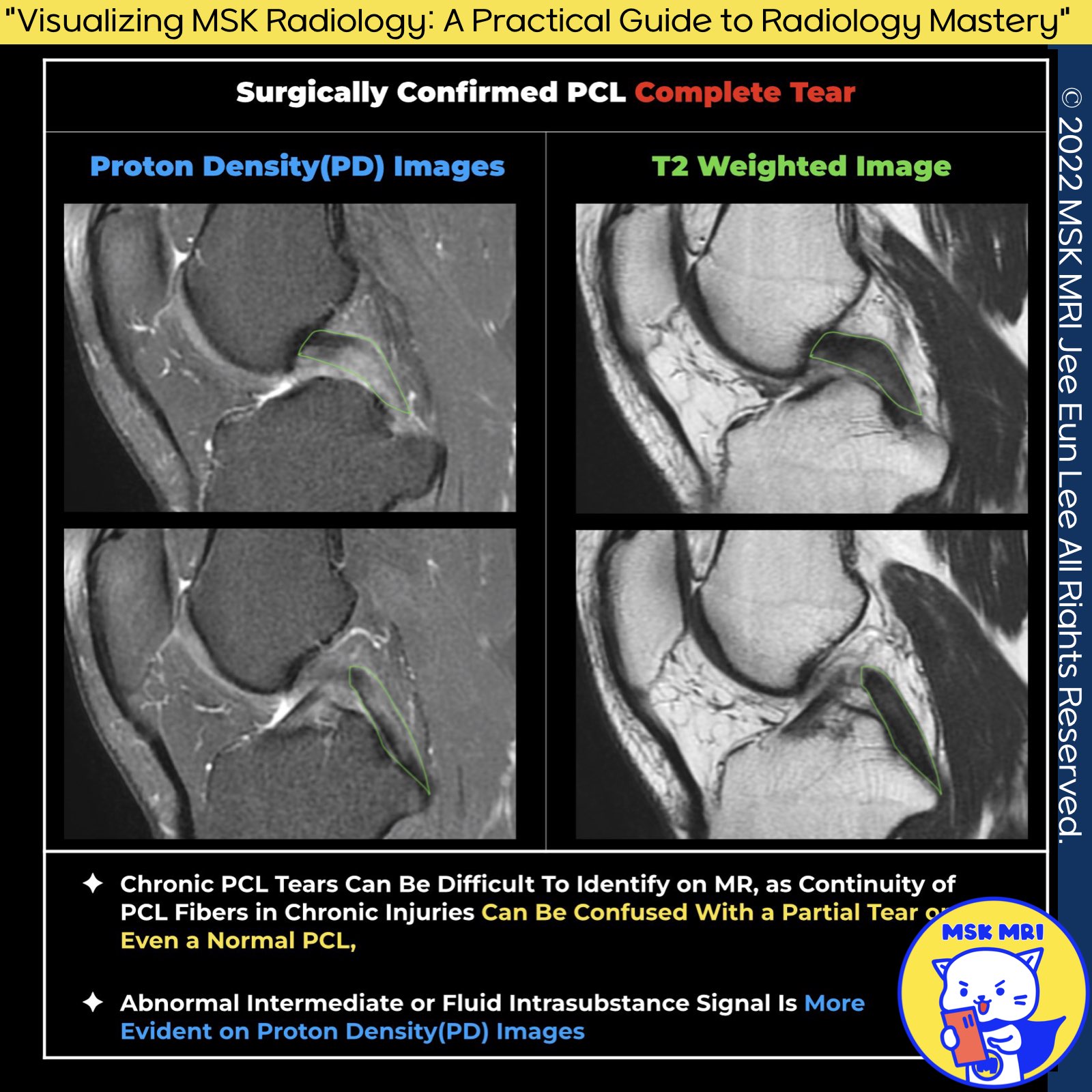
2️⃣: The Role of Proton Density Imaging
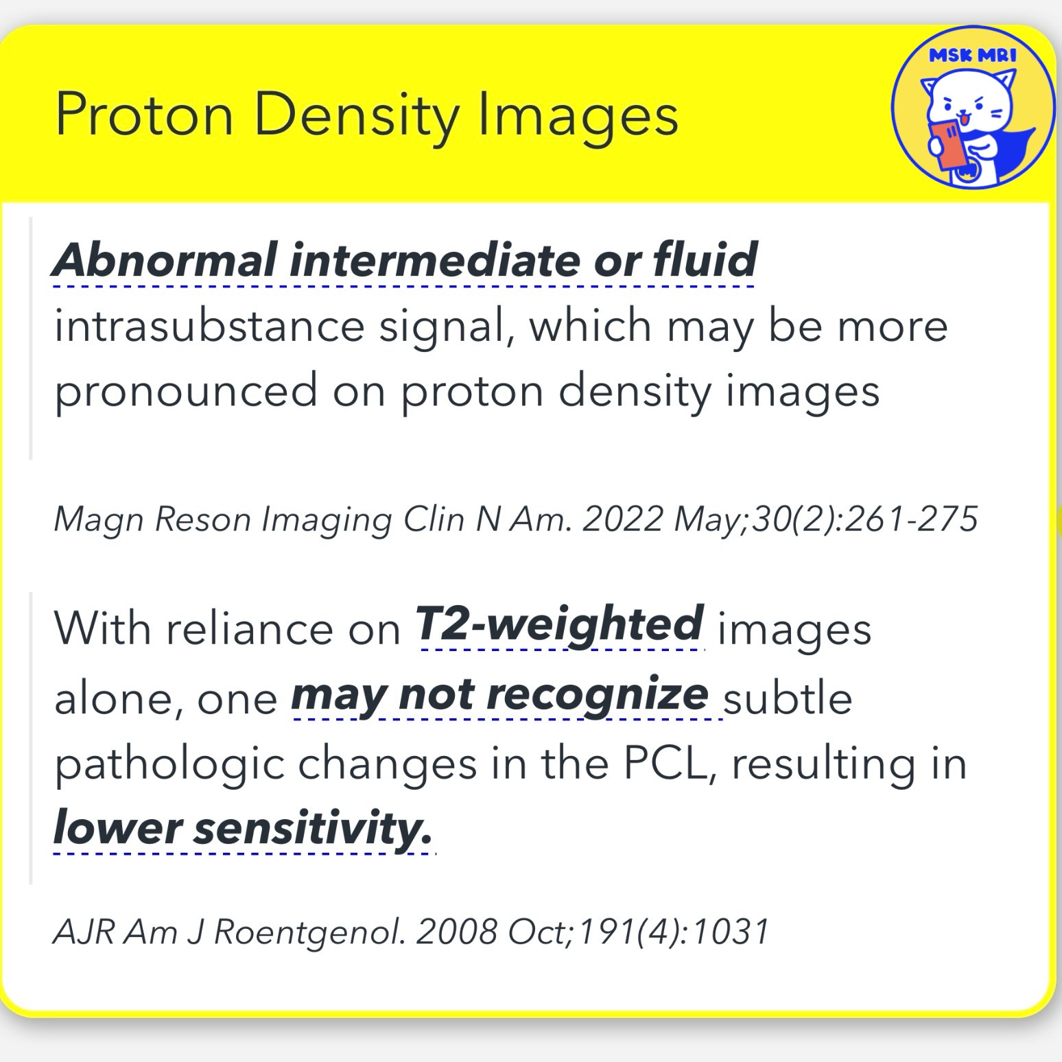
- Proton density imaging, particularly with fat suppression, is crucial for highlighting subtle pathological changes within the PCL. T
- his technique often reveals increased intraligamentous signal intensity that might not be evident on T2-weighted images.
- For instance, in a case where the PCL diameter and contour appeared normal on T2WI, the proton density sequence unveiled significant abnormalities leading to a correct diagnosis of
3️⃣: The Value of Stress Radiography
- In cases where MRI results are equivocal, stress radiographs provide an objective method to evaluate the structural integrity of the PCL.
- By quantifying the extent of posterior tibial translation, this technique helps distinguish between partial and complete tears. Measurements show that less than 8mm of translation usually indicates a partial tear, 8-12mm a complete tear, and more than 12mm typically suggests a combined complete tear involving the posterolateral corner.
Magn Reson Imaging Clin N Am 22 (2014) 557–580
Magn Reson Imaging Clin N Am. 2022 May;30(2):261-275
AJR Am J Roentgenol. 2008 Oct;191(4):1031
Am J Sports Med. 2015 Dec;43(12):3077-92

'✅ Knee MRI Mastery > Chap 2.ACL and PCL' 카테고리의 다른 글
| (Fig 2-E.26) PCL Repair (0) | 2024.04.28 |
|---|---|
| (Fig 2-E.25) Multiligamentous Injuries Associated with PCL Injuries (0) | 2024.04.28 |
| (Fig 2-E.23) Chronic Complete Tear of the PCL - Part 1 (2) | 2024.04.28 |
| (Fig 2-E.22) Posterior Tibial Translation (0) | 2024.04.28 |
| (Fig 2-E.21) Radiographic Assessment of Posterior Knee Laxity (0) | 2024.04.27 |