Click the link to purchase on Amazon 🎉📚
==============================================
🎥 Check Out All Videos at Once! 📺
👉 Visit Visualizing MSK Blog to explore a wide range of videos! 🩻
https://visualizingmsk.blogspot.com/?view=magazine
📚 You can also find them on MSK MRI Blog and Naver Blog! 📖
https://www.instagram.com/msk_mri/
Click now to stay updated with the latest content! 🔍✨
==============================================
📌Anatomy of the Medial Side of the Knee: Two Approaches
✅ Warren et al.'s Approach:
Initially, Warren et al. divided the medial side into three layers:
- Layer I: Deep fascia
- Layer II: Superficial MCL
- Layer III: Joint capsule and deep MCL
✅ Robinson et al.'s Approach:
A more recent description by Robinson et al. divides the medial side into thirds:
- Anterior Third: From the medial border of the patellar tendon to the anterior border of the longitudinal fibers of the superficial MCL
- Middle Third: Composed of the width of the longitudinal fibers of the MCL
- Posterior Third: Encompassing the Posteromedial Corner (PMC), located between the posterior margin of the superficial MCL's longitudinal fibers and the medial border of the PCL
✅ Importance of Posteromedial Corner (PMC) Injuries:
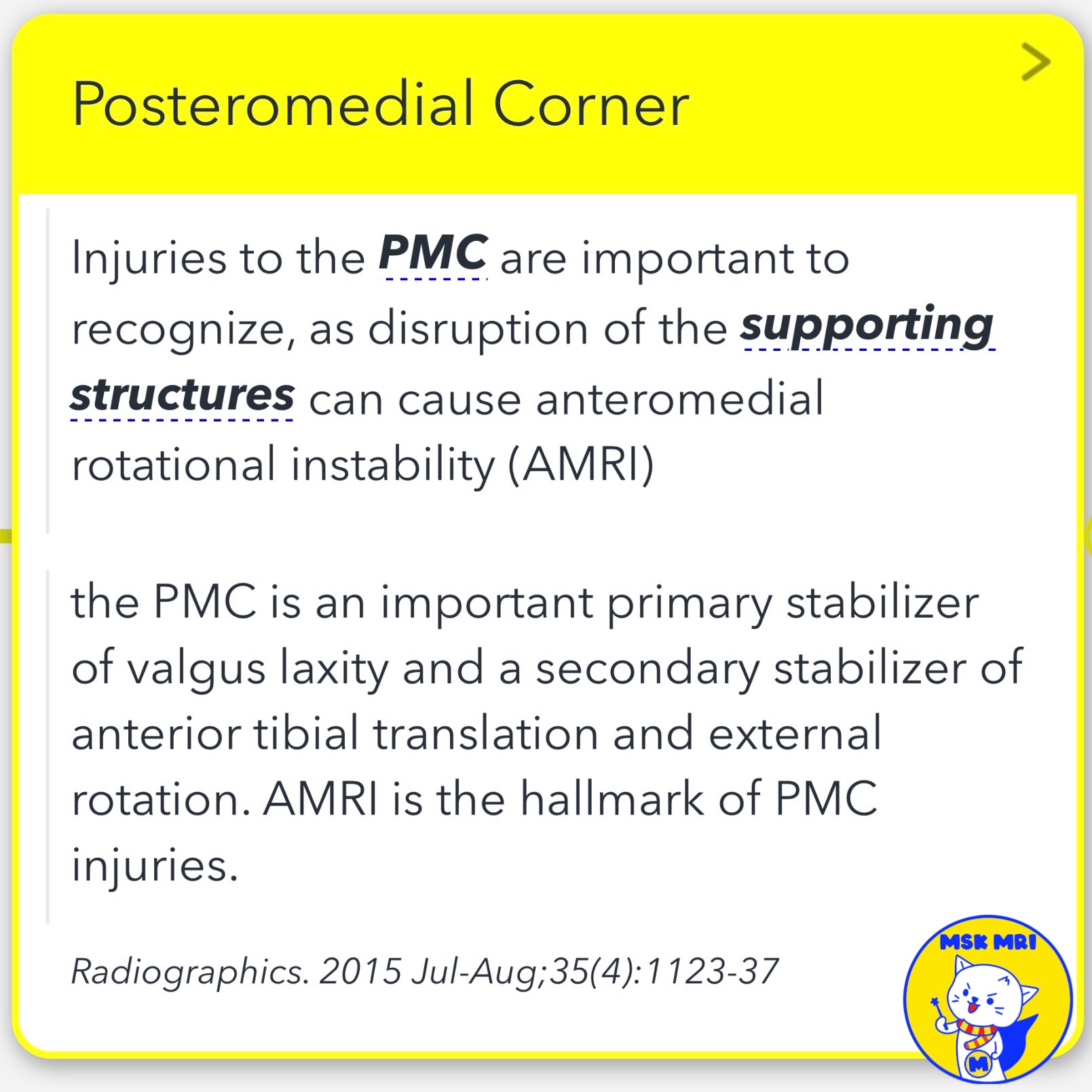
➡️ Injuries to the PMC are crucial to recognize, as they can lead to anteromedial rotational instability (AMRI).
➡️ The PMC serves as a primary stabilizer for valgus laxity and a secondary stabilizer for anterior tibial translation and external rotation, with AMRI being the hallmark of PMC injuries.
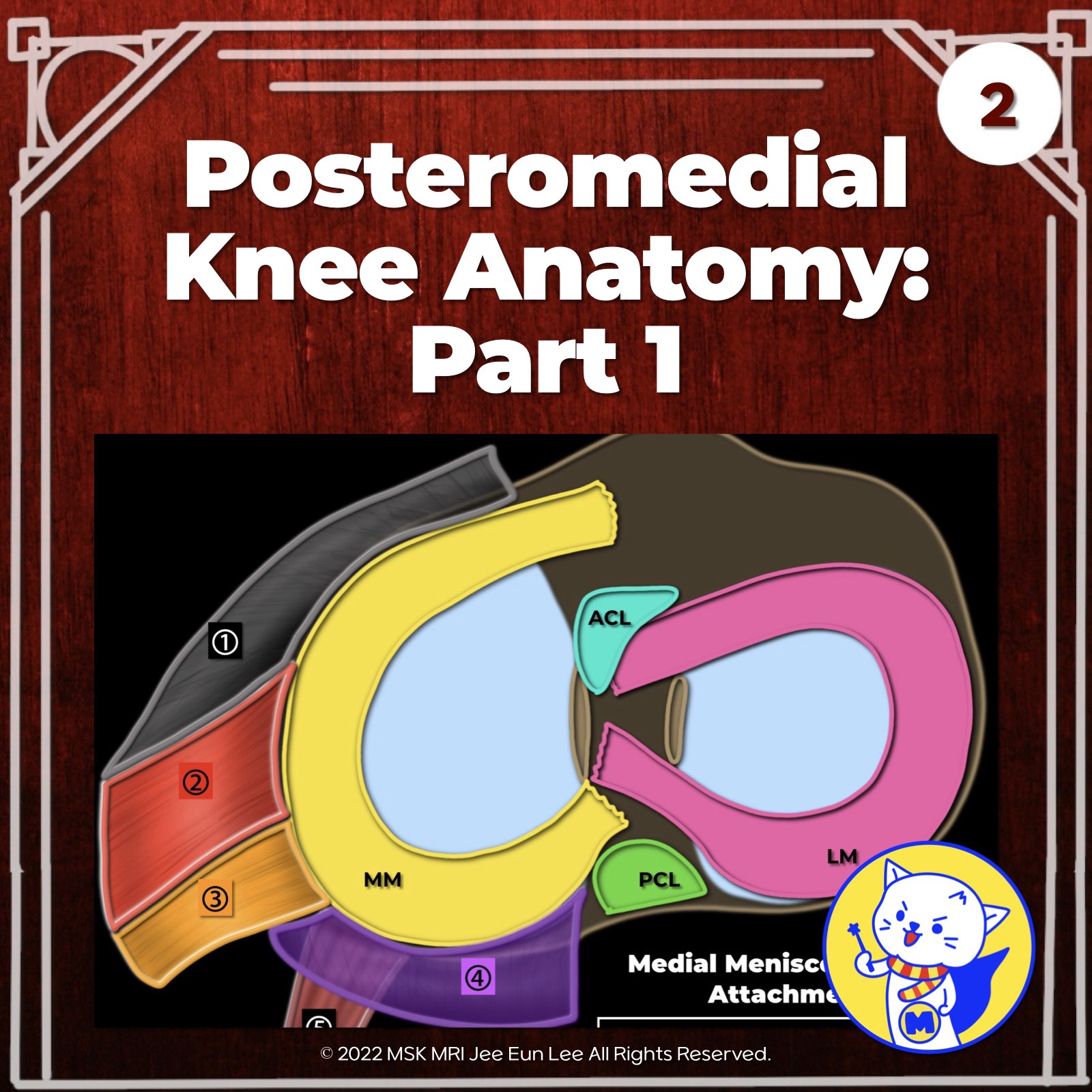
1️⃣Semimembranosus muscle has anterior, inferior, direct, oblique popliteal ligament, and capsular expansions. The anterior arm connects to the tibia, passing under the posterior oblique ligament. There's also a short arm attaching to the coronary ligament of the medial meniscus.
2️⃣ The oblique popliteal ligament (OPL) extends obliquely from the semimembranosus muscle-tendon complex to the lateral head of the gastrocnemius muscle and plantaris muscle, closely associated with the arcuate ligament.
3️⃣The posterior oblique ligament (POL) is part of layer II. It connects the superficial medial collateral ligament to the posterior meniscocapsular attachment and blends with the joint capsule posteriorly.
4️⃣ The meniscocapsular ligament and meniscotibial ligament attach the posterior horn of the medial meniscus to the joint capsule and tibia, respectively.
5️⃣ The posterior horn of the medial meniscus
"Visualizing MSK Radiology: A Practical Guide to Radiology Mastery"
© 2022 MSK MRI Jee Eun Lee All Rights Reserved.
No unauthorized reproduction, redistribution, or use for AI training.
#MCL, #Medialknee, #POL, #OPL, #Patellarretinaculum, #kneeanatomy, #anatomyknee #PMC, #Semimembranosus, #AMRI,