==============================================
⬇️✨⬇️🎉⬇️🔥⬇️📚⬇️
Click the link to purchase on Amazon 🎉📚
==============================================
🎥 Check Out All Videos at Once! 📺
👉 Visit Visualizing MSK Blog to explore a wide range of videos! 🩻
https://visualizingmsk.blogspot.com/?view=magazine
📚 You can also find them on MSK MRI Blog and Naver Blog! 📖
https://www.instagram.com/msk_mri/
Click now to stay updated with the latest content! 🔍✨
==============================================
📌 Superficial Medial Collateral Ligament (sMCL)
- Band-like structure with one femoral and two tibial attachments
- Measures around 1.5 cm anteroposterior and 9.5 cm long
- Femoral attachment: 3.2 mm proximal, 4.8 mm posterior to medial epicondyle
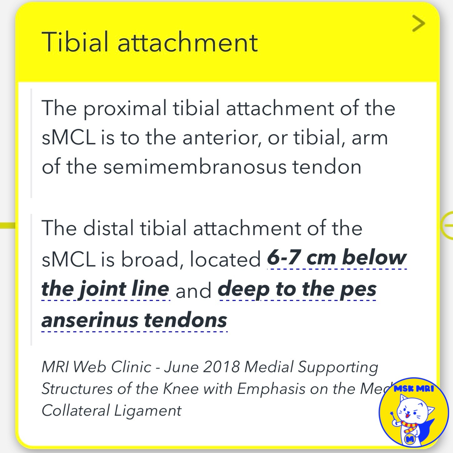
- Proximal tibial attachment: Blends with distal semimembranosus tendon sheath, 10-12 mm distal to joint line
- Distal tibial attachment: Broad, 6-7 cm below joint line, deep to pes anserinus tendons
✅ Pes Anserinus Tendons
- Sartorius, gracilis, and semitendinosus tendons attach to anteromedial tibia
✅ Importance of sMCL Anatomy
- Recognize subtle Stener-like lesions
- Interposed pes tendons can prevent sMCL healing, leading to chronic valgus instability
📌 Deep Medial Collateral Ligament (dMCL)
- Thickening of medial joint capsule, associated with medial meniscus
- Meniscofemoral portion: Distal femur to medial meniscus
- Meniscotibial portion: Medial meniscus to proximal tibia
- Posterior border blends with central arm of posterior oblique ligament
- Part of Layer 3 (dMCL, posterior oblique ligament, oblique popliteal ligament)
- Distinguishable from sMCL anteriorly, blends posteriorly
✅ MCL Bursitis
- MCL bursa between dMCL and sMCL reduces friction during flexion
- Delineates sMCL and dMCL more clearly
"Visualizing MSK Radiology: A Practical Guide to Radiology Mastery"
© 2022 MSK MRI Jee Eun Lee All Rights Reserved.
No unauthorized reproduction, redistribution, or use for AI training.
#MCL, #Medialknee, #POL, #kneeanatomy, #anatomyknee #sMCL, #dMCL