Click the link to purchase on Amazon 🎉📚
==============================================
🎥 Check Out All Videos at Once! 📺
👉 Visit Visualizing MSK Blog to explore a wide range of videos! 🩻
https://visualizingmsk.blogspot.com/?view=magazine
📚 You can also find them on MSK MRI Blog and Naver Blog! 📖
https://www.instagram.com/msk_mri/
Click now to stay updated with the latest content! 🔍✨
==============================================
📌 Attachments of the medial collateral and posterior oblique ligaments
1️⃣ sMCL-femoral attachment
Historically reported to be attached to the medial epicondyle (ME)
LaPrade et al.: Recent publications suggest the attachment is 3.2mm proximal and 4.8mm posterior to the ME
Imperial College London: demonstrated that the sMCL covers the ME, with the attachment centered 1-2mm proximal
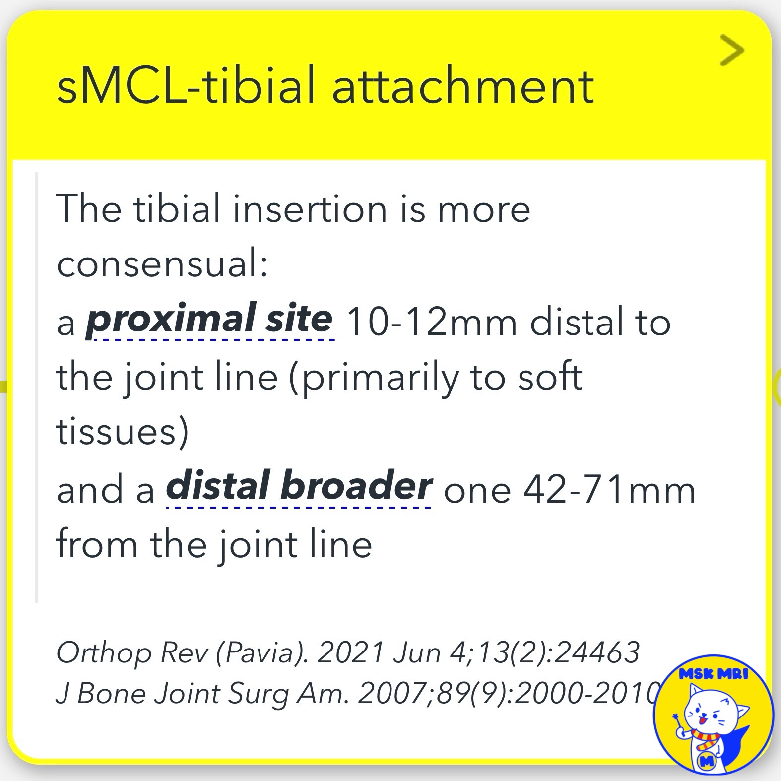
2️⃣ sMCL-tibial attachment
A proximal site 10-12mm distal to the joint line (primarily to soft tissues)
A broader attachment 42-71mm from the joint line
3️⃣ dMCL-femoral attachment
An important independent stabilizer, despite being adherent to the articular capsule
Proximal to the sMCL (6mm distal and 5mm posterior to the medial epicondyle)
15-17mm above the femoral articular cartilage
4️⃣ dMCL-tibial attachment
Runs distally from posterior to anterior
Ends in a fan-wide tibial attachment around 8mm distal to the joint line
Has two major expansions to the meniscus: meniscofemoral and meniscotibial ligament
5️⃣ POL-femoral attachment
Composed of obliquely oriented fibers extending from the distal tendon of the SM toward the femur
Anteriorly blends with the posterior margin of the sMCL and posteriorly reinforces the articular capsule
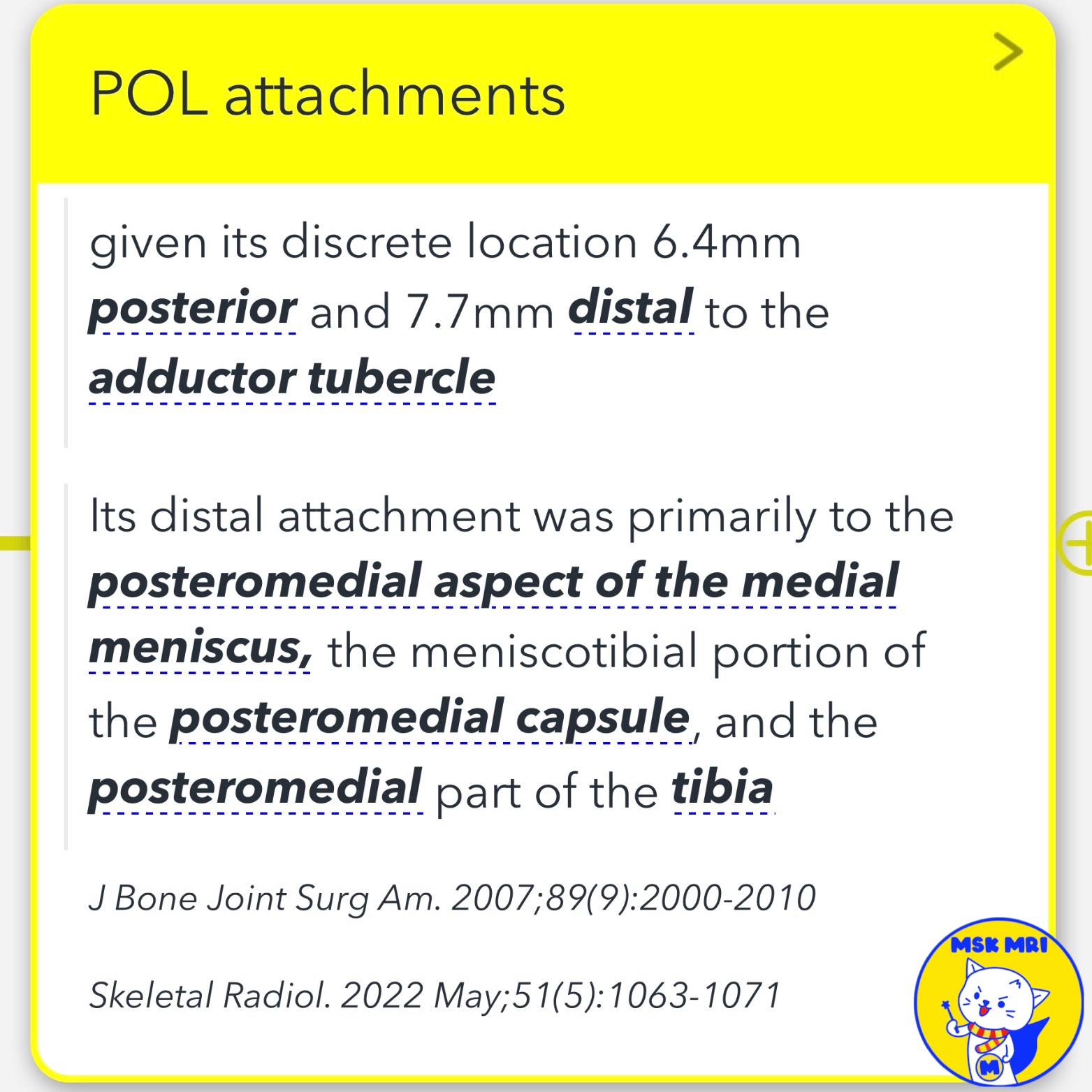
Accepted as an individual structure, located 6.4mm posterior and 7.7mm distal to the adductor tubercle
6️⃣ POL-distal attachment (central arm)
Distal attachment primarily to the posteromedial aspect of the medial meniscus, the meniscotibial portion of the posteromedial capsule, and the posteromedial part of the tibia
"Visualizing MSK Radiology: A Practical Guide to Radiology Mastery"
© 2022 MSK MRI Jee Eun Lee All Rights Reserved.
No unauthorized reproduction, redistribution, or use for AI training.
#MCL, #Medialknee, #POL, #OPL, #kneeanatomy, #anatomyknee #POL,
'✅ Knee MRI Mastery > Chap 3.Collateral Ligaments' 카테고리의 다른 글
| (Fig 3-A.06) Medial Collateral Ligament Anatomy, Knee MRI, coronal and axial images (0) | 2024.05.04 |
|---|---|
| (Fig 3-A.05) Medial Collateral Ligament Anatomy, Knee MRI, coronal and axial images (0) | 2024.05.04 |
| (Fig 3-A.03) Posteromedial Knee Anatomy: Part 2 (0) | 2024.05.04 |
| (Fig 3-A.02) Posteromedial Knee Anatomy: Part 1 (0) | 2024.05.03 |
| (Fig 3-A.01) Three-Layer Approach to Medial Knee (0) | 2024.05.02 |


