https://youtu.be/_qSjjjH5v6c?si=vYrQsBic3OSHrdd3
Click the link to purchase on Amazon 🎉📚
==============================================
🎥 Check Out All Videos at Once! 📺
👉 Visit Visualizing MSK Blog to explore a wide range of videos! 🩻
https://visualizingmsk.blogspot.com/?view=magazine
📚 You can also find them on MSK MRI Blog and Naver Blog! 📖
https://www.instagram.com/msk_mri/
Click now to stay updated with the latest content! 🔍✨
==============================================
CASE STUDY
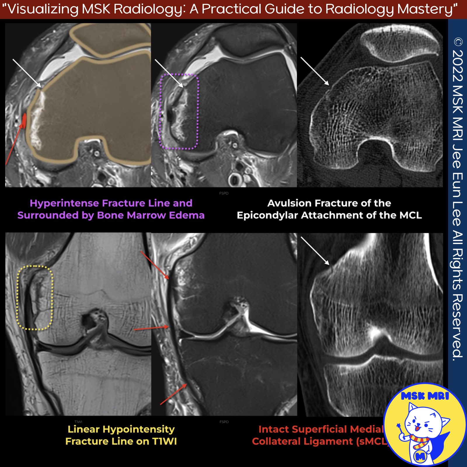
✅Avulsion fracture at the medial collateral ligament's epicondylar attachment.
✅ Key findings on imaging include:
- Avulsion fracture of the MCL visible, without displacement
- Continuity of the fracture fragment with the MCL on MRI
- Associated extracapsular hemorrhage appearing hyperintense
- Fracture line is hyperintense surrounded by bone marrow edema, making it less apparent on fat-suppressed images
- Fracture line appears as linear hypointensity on T1-weighted MRI
✅Note
It's important to assess for associated injuries like bone marrow edema, injury to posteromedial corner structures, deep MCL, medial meniscus, and cruciate ligaments which can accompany this type of avulsion fracture.
Emerg Radiol. 2012 Dec;19(6):489-98
"Visualizing MSK Radiology: A Practical Guide to Radiology Mastery"
© 2022 MSK MRI Jee Eun Lee All Rights Reserved.
No unauthorized reproduction, redistribution, or use for AI training.
#MCL, #sMCL, #MCLinjury, #MCLavulsion, #avulsionfracture, #fracture, #femurfracture, #Valgusinjury,