Click the link to purchase on Amazon 🎉📚
==============================================
🎥 Check Out All Videos at Once! 📺
👉 Visit Visualizing MSK Blog to explore a wide range of videos! 🩻
https://visualizingmsk.blogspot.com/?view=magazine
📚 You can also find them on MSK MRI Blog and Naver Blog! 📖
https://www.instagram.com/msk_mri/
Click now to stay updated with the latest content! 🔍✨
==============================================
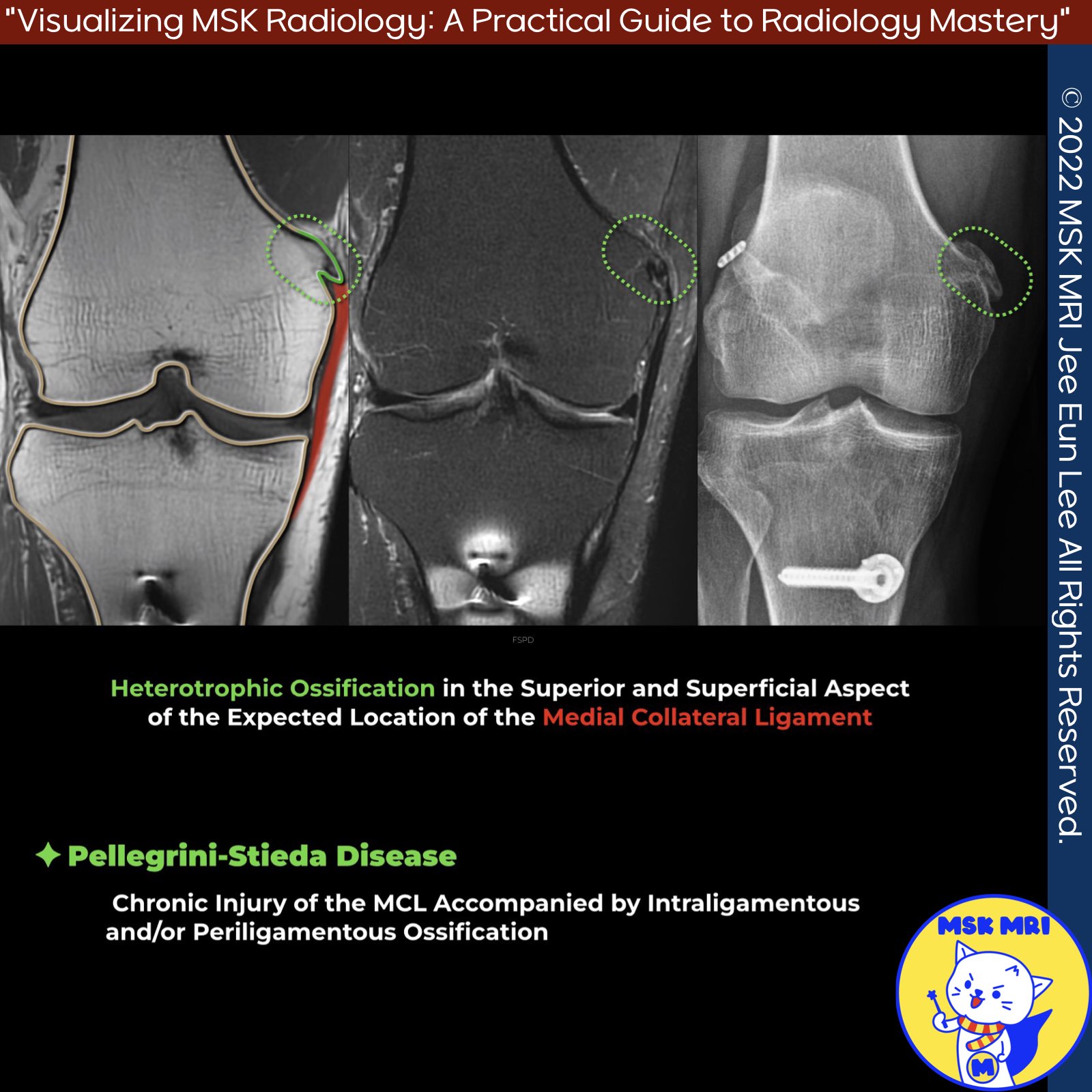
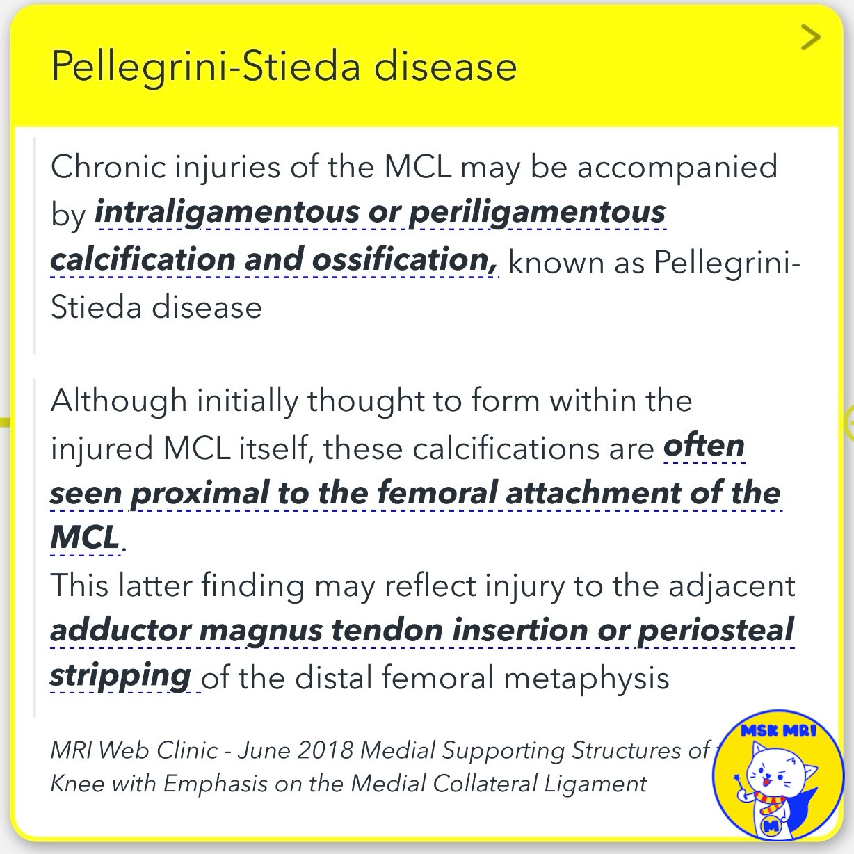
📌 Pellegrini-Stieda disease
: refers to intraligamentous or periligamentous calcification and ossification that can occur with chronic injuries of the medial collateral ligament (MCL).
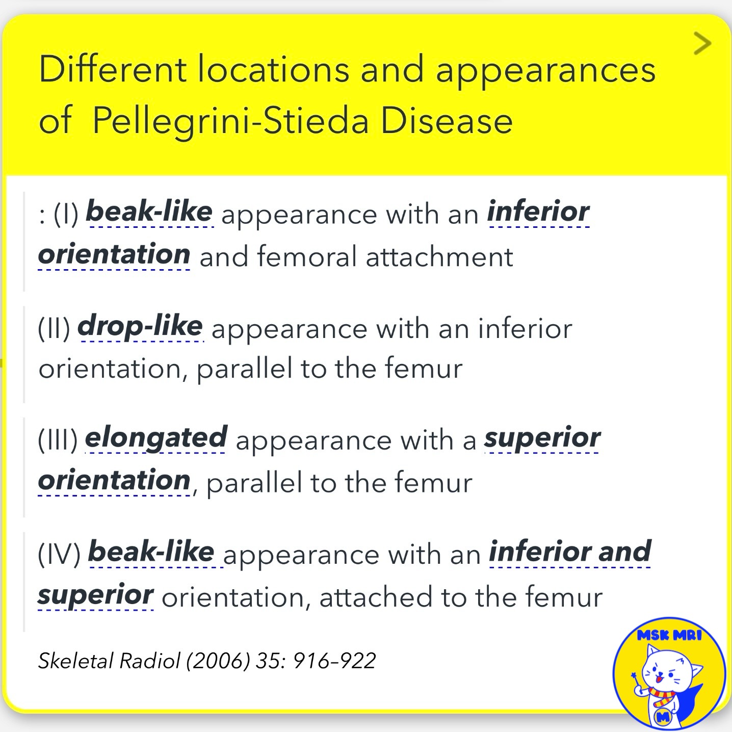
✅ Schematic Representation
This condition can present with different appearances and locations around the MCL, as depicted in the provided schematic:
I. Beak-like appearance with inferior orientation and femoral attachment
II. Drop-like appearance with inferior orientation, parallel to the femur III. Elongated appearance with superior orientation, parallel to the femur
IV. Beak-like appearance with both inferior and superior orientation, attached to the femur
✅ While most patients with Pellegrini-Stieda disease are asymptomatic, a small proportion may experience medial knee pain.
Skeletal Radiol (2006) 35: 916–922
MRI Web Clinic - June 2018 Medial Supporting Structures of the Knee with Emphasis on the Medial Collateral Ligament
"Visualizing MSK Radiology: A Practical Guide to Radiology Mastery"
© 2022 MSK MRI Jee Eun Lee All Rights Reserved.
No unauthorized reproduction, redistribution, or use for AI training.
#MCL, #sMCL, #MCLinjury, #MCLtear, #MPFL, #chronicMCLinjury, #Pellegrini, #Stieda, #Stiedadisease,


'✅ Knee MRI Mastery > Chap 3.Collateral Ligaments' 카테고리의 다른 글
| (Fig 3-A.20) Superficial and Deep MCL Anatomy (1) | 2024.05.08 |
|---|---|
| (Fig 3-A.19) Pellegrini-Stieda Disease_ Part 2 (0) | 2024.05.08 |
| (Fig 3-A.17) Subacute to Chronic MCL Injury (0) | 2024.05.07 |
| (Fig 3-A.16) MCL Avulsion fracture (0) | 2024.05.07 |
| (Fig 3-A.14) Intra-Articular Entrapment of the MCL: Part 1, medial patellofemoral ligament, POL tear (0) | 2024.05.06 |