Click the link to purchase on Amazon 🎉📚
==============================================
🎥 Check Out All Videos at Once! 📺
👉 Visit Visualizing MSK Blog to explore a wide range of videos! 🩻
https://visualizingmsk.blogspot.com/?view=magazine
📚 You can also find them on MSK MRI Blog and Naver Blog! 📖
https://www.instagram.com/msk_mri/
Click now to stay updated with the latest content! 🔍✨
==============================================
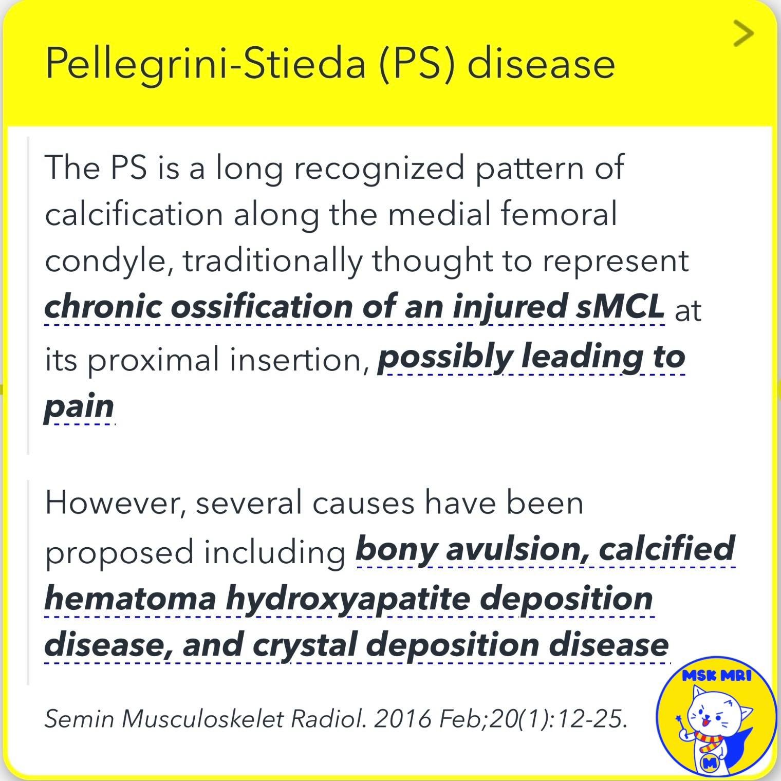
✅Main imaging findings of Pellegrini-Stieda Disease
- A large ossification adjacent to the medial femoral epicondyle with marrow fat signal or uniform fat suppression
- Intraligamentous and periligamentous ossification of the MCL, with thickening and calcification at the proximal insertion and mid-substance
- Potential ossification proximal to the MCL, suggesting injury to the adductor magnus tendon insertion, heterotopic ossification, or periosteal stripping.
MRI Web Clinic - June 2018 Medial Supporting Structures of the Knee with Emphasis on the Medial Collateral Ligament
Stoller's Orthopaedics and Sports Medicine:
The Knee: Includes Stoller Lecture Videos and Stoller Notes
"Visualizing MSK Radiology: A Practical Guide to Radiology Mastery"
© 2022 MSK MRI Jee Eun Lee All Rights Reserved.
No unauthorized reproduction, redistribution, or use for AI training.
#MCL, #sMCL, #MCLinjury, #MCLtear, #MPFL, #chronicMCLinjury, #Pellegrini, #Stieda, #Stiedadisease,


'✅ Knee MRI Mastery > Chap 3.Collateral Ligaments' 카테고리의 다른 글
| (Fig 3-A.21) Meniscofemoral Ligament Tear (0) | 2024.05.08 |
|---|---|
| (Fig 3-A.20) Superficial and Deep MCL Anatomy (1) | 2024.05.08 |
| (Fig 3-A.18) Pellegrini-Stieda Disease: Part 1 (0) | 2024.05.07 |
| (Fig 3-A.17) Subacute to Chronic MCL Injury (0) | 2024.05.07 |
| (Fig 3-A.16) MCL Avulsion fracture (0) | 2024.05.07 |