Click the link to purchase on Amazon 🎉📚
==============================================
🎥 Check Out All Videos at Once! 📺
👉 Visit Visualizing MSK Blog to explore a wide range of videos! 🩻
https://visualizingmsk.blogspot.com/?view=magazine
📚 You can also find them on MSK MRI Blog and Naver Blog! 📖
https://www.instagram.com/msk_mri/
Click now to stay updated with the latest content! 🔍✨
==============================================
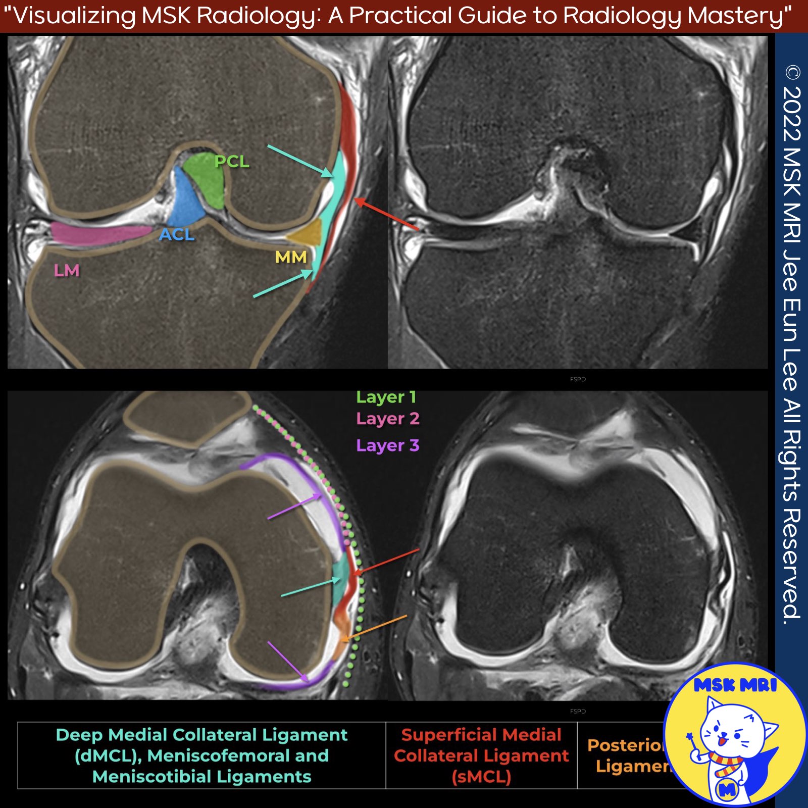
📌Deep Medial Collateral Ligament
- The deep medial collateral ligament (dMCL) is an essential independent stabilizer of the knee joint, adhering to the articular capsule.
- Rupture of the dMCL can clinically mimic a meniscal tear, highlighting the importance of recognizing and properly evaluating this structure during knee examinations and imaging studies.
✅Anatomy Orientation and Attachments
- Recent findings have revealed that the dMCL has a distinctive fan-shaped orientation, extending antero-distally towards a 22 mm wide tibial attachment located approximately 8 mm distal to the joint line.
- The dMCL is a thickening of the medial joint capsule.
- It has a broad, firm attachment to the midbody of the medial meniscus, situated between the meniscofemoral attachment of the posterior oblique ligament (POL) and the anteromedial capsule.
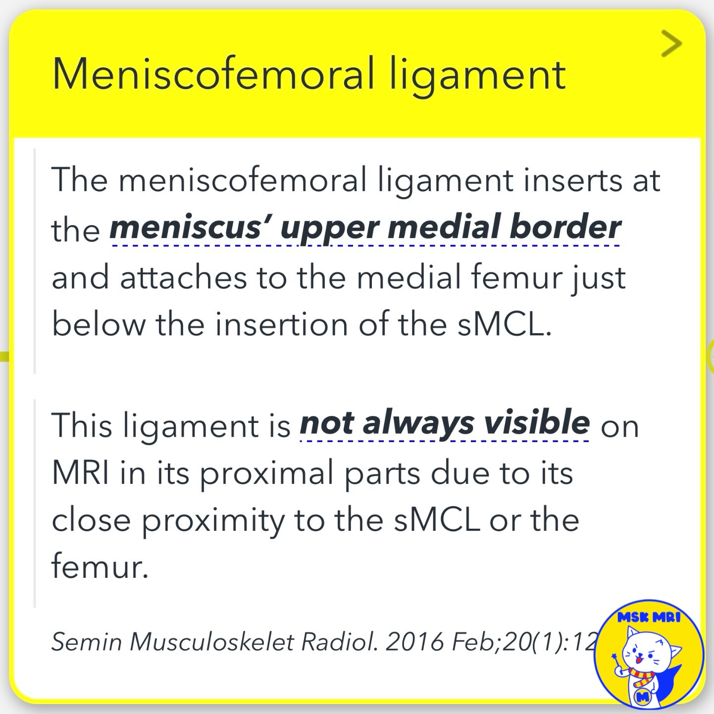
✅ Meniscofemoral ligament:
- Inserts at the meniscus' upper medial border
- Attaches to the medial femur just below the insertion of the superficial MCL (sMCL)
- May not be visible on MRI due to its proximity to the sMCL or femur
✅ Meniscotibial ligament:
- Inserts at the meniscus' lower medial border
- Has a short course to its attachment at the medial corner of the tibia, immediately below the joint line
Knee Surgery, Sports Traumatology, Arthroscopy (2020) 28:3709–3719
Orthop Rev (Pavia). 2021 Jun 4;13(2):24463
The American Journal of Sports Medicine 2019;47(2):372–378
Semin Musculoskelet Radiol. 2016 Feb;20(1):12-25
"Visualizing MSK Radiology: A Practical Guide to Radiology Mastery"
© 2022 MSK MRI Jee Eun Lee All Rights Reserved.
No unauthorized reproduction, redistribution, or use for AI training.
#MCL, #Medialknee, #POL, #OPL, #kneeanatomy, #anatomyknee #POL,
In summary, the dMCL is a crucial independent stabilizer of the knee joint, with distinct attachments to the tibial plateau, medial meniscus, and associated meniscofemoral and meniscotibial ligaments. Its strategic location and connections underscore its significance in maintaining knee stability and proper function.


'✅ Knee MRI Mastery > Chap 3.Collateral Ligaments' 카테고리의 다른 글
| (Fig 3-A.22) Meniscofemoral Ligament Avulsion Fracture (0) | 2024.05.09 |
|---|---|
| (Fig 3-A.21) Meniscofemoral Ligament Tear (0) | 2024.05.08 |
| (Fig 3-A.19) Pellegrini-Stieda Disease_ Part 2 (0) | 2024.05.08 |
| (Fig 3-A.18) Pellegrini-Stieda Disease: Part 1 (0) | 2024.05.07 |
| (Fig 3-A.17) Subacute to Chronic MCL Injury (0) | 2024.05.07 |