Click the link to purchase on Amazon 🎉📚
==============================================
🎥 Check Out All Videos at Once! 📺
👉 Visit Visualizing MSK Blog to explore a wide range of videos! 🩻
https://visualizingmsk.blogspot.com/?view=magazine
📚 You can also find them on MSK MRI Blog and Naver Blog! 📖
https://www.instagram.com/msk_mri/
Click now to stay updated with the latest content! 🔍✨
==============================================
출처: https://mskmri.tistory.com/1056 [MSK MRI:티스토리]
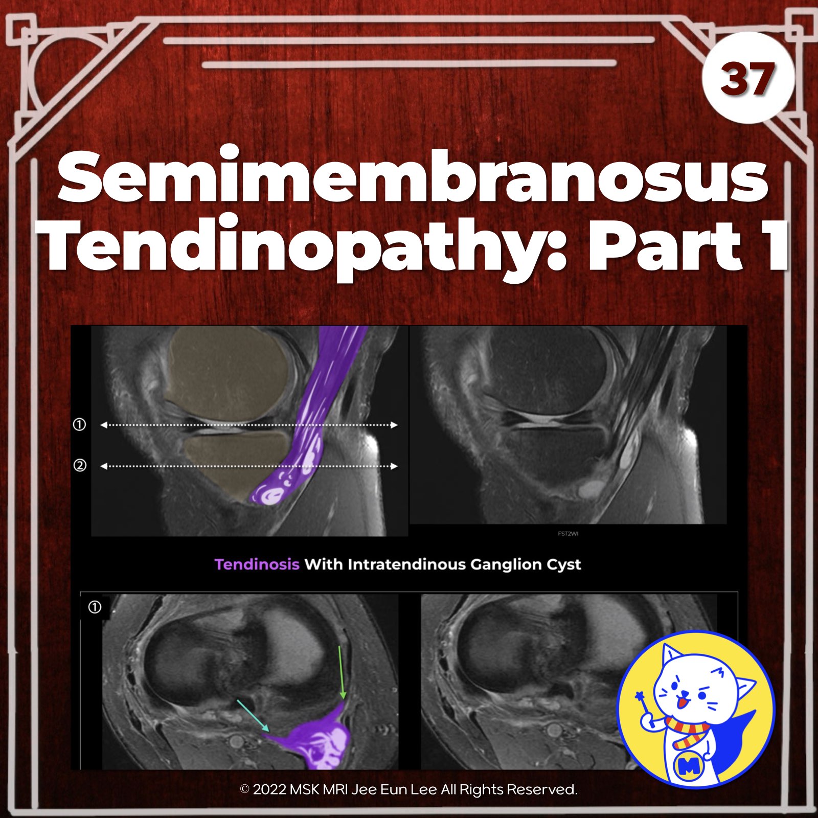
✅ Semimembranosus Insertion Injuries
Injuries at the distal semimembranosus insertion are observed in up to 70% of posteromedial corner injuries.
These can range from avulsion fractures at its tibial attachment to partial or complete tendon tears, as well as tendinosis.
✅ Pathophysiology of Tendinosis
Tendinosis in this context is often the result of chronic stress, leading to thickening of the tendon insertion.
This alteration in the tendon’s structure compromises its function and increases the susceptibility to further injury.
✅ Intratendinous Ganglion Cyst of the Semimembranosus Tendon
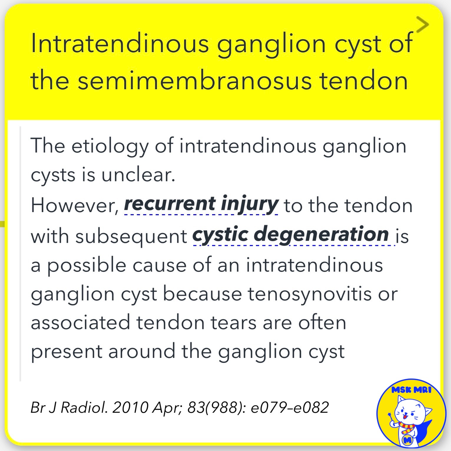
The etiology of intratendinous ganglion cysts remains somewhat unclear.
However, it is postulated that recurrent injury leading to cystic degeneration might be a contributing factor.
These cysts often coexist with conditions like tenosynovitis or associated tendon tears, which exacerbate the complexity of the clinical presentation.
Radiol Clin North Am. 2013 May;51(3):413-32
Br J Radiol. 2010 Apr; 83(988): e079–e082
"Visualizing MSK Radiology: A Practical Guide to Radiology Mastery"
© 2022 MSK MRI Jee Eun Lee All Rights Reserved.
No unauthorized reproduction, redistribution, or use for AI training.
#semimembranosus, #kneeMRI, #posteromedialcorner, #Kneeanatomy, #semimembranosustendon, #tendinosis #ganglioncyst, #intratendinosus
'✅ Knee MRI Mastery > Chap 3.Collateral Ligaments' 카테고리의 다른 글
| (Fig 3-A.39) Fat Accumulation in Semimembranosus Tendon (0) | 2024.05.12 |
|---|---|
| (Fig 3-A.38) Semimembranosus Tendinopathy: Part 2 (0) | 2024.05.12 |
| (Fig 3-A.36) Magic Angle Artifact in Semimembranosus Tendon (0) | 2024.05.12 |
| (Fig 3-A.35) Semimembranosus Arms with Bursitis: Anterior, Direct, Inferior (0) | 2024.05.12 |
| (Fig 3-A.34) Anterior and Direct Semimembranosus Arms (0) | 2024.05.12 |


