Click the link to purchase on Amazon 🎉📚
==============================================
🎥 Check Out All Videos at Once! 📺
👉 Visit Visualizing MSK Blog to explore a wide range of videos! 🩻
https://visualizingmsk.blogspot.com/?view=magazine
📚 You can also find them on MSK MRI Blog and Naver Blog! 📖
https://www.instagram.com/msk_mri/
Click now to stay updated with the latest content! 🔍✨
==============================================
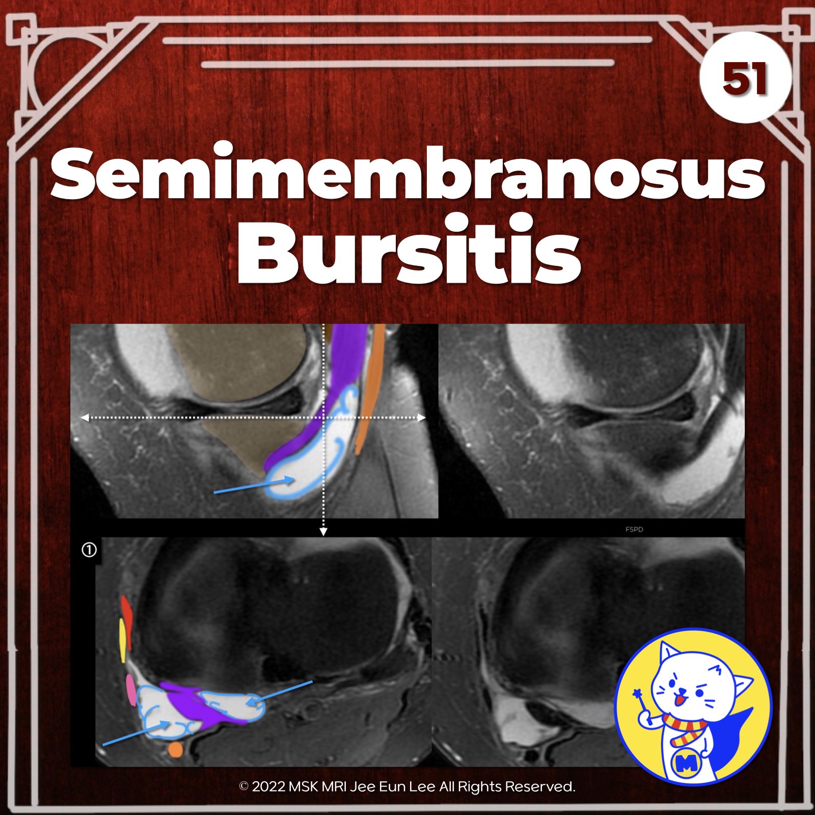
📌Semimembranosus Bursa:
- Horseshoe-shaped synovial bursa around the semimembranosus tendon near posteromedial tibial rim
- Inflamed/distended bursa presents as a synovial-lined space wrapping the tendon
- Demonstrates characteristic reverse 'J' shape or 'inverted-U' shape on imaging
✅Direct Arm of Semimembranosus Tendon
- Principal attachment of the semimembranosus tendon
- Attaches to posteromedial aspect of tibia, just distal to the joint line
- Forms a C-shaped 5-mm-thick band with the anterior arm
✅Anterior Arm of Semimembranosus Tendon
- Seen on peripheral medial sagittal images, curving anteriorly
- Appears as a round hypointense structure on coronal images, adjacent to medial tibia, passing under the MCL
Semin Musculoskelet Radiol. 2016 Feb;20(1):12-25
MRI Web Clinic - June 2018 Medial Supporting Structures of the Knee with Emphasis on the Medial Collateral Ligament
Radiographics. 2015 Jul-Aug;35(4):1123-37
"Visualizing MSK Radiology: A Practical Guide to Radiology Mastery"
© 2022 MSK MRI Jee Eun Lee All Rights Reserved.
No unauthorized reproduction, redistribution, or use for AI training.
#PMC, #POL, #semimembranosus, #kneeMRI, #posteromedialcorner, #Kneeanatomy, #semimembranosustendon, #bursitis,


'✅ Knee MRI Mastery > Chap 3.Collateral Ligaments' 카테고리의 다른 글
| (Fig 3-A.53) MCL Bursitis ⎜Distinguishing from Grade I MCL Injury (0) | 2024.05.15 |
|---|---|
| (Fig 3-A.52) Semimembranosus-Gastrocnemius Bursa: Baker Cyst, Synovial Osteochondromatosis (0) | 2024.05.15 |
| (Fig 3-A.50) Pes Anserine Bursitis (0) | 2024.05.14 |
| (Fig 3-A.48) Osteomeniscal Impingement (0) | 2024.05.14 |
| (Fig 3-A.47) Extra-Capsular Medial Tibial Crest Friction (0) | 2024.05.14 |