Click the link to purchase on Amazon 🎉📚
==============================================
🎥 Check Out All Videos at Once! 📺
👉 Visit Visualizing MSK Blog to explore a wide range of videos! 🩻
https://visualizingmsk.blogspot.com/?view=magazine
📚 You can also find them on MSK MRI Blog and Naver Blog! 📖
https://www.instagram.com/msk_mri/
Click now to stay updated with the latest content! 🔍✨
==============================================
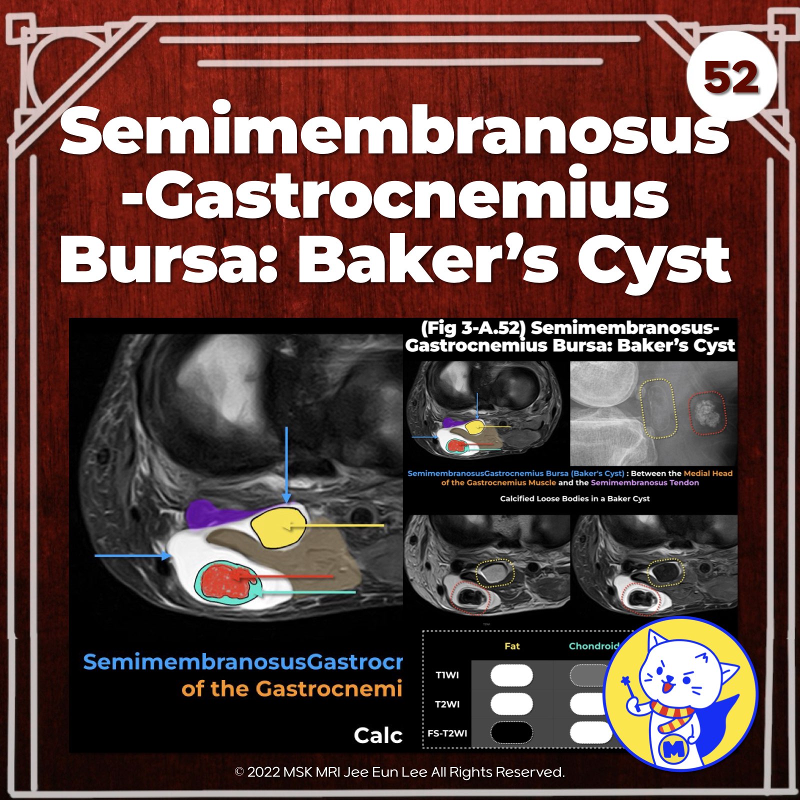
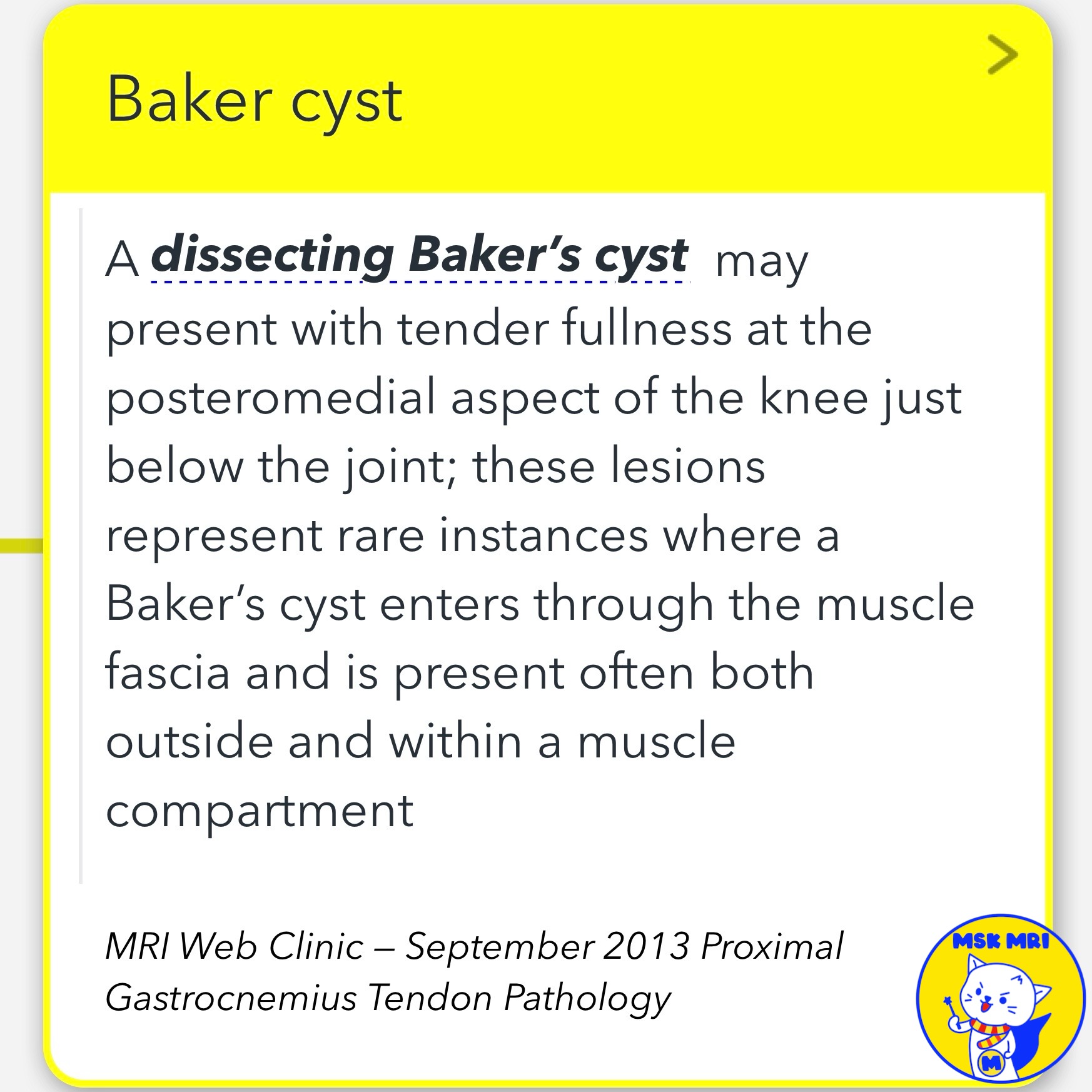
📌 Semimembranosus-Gastrocnemius Bursa: Baker Cyst
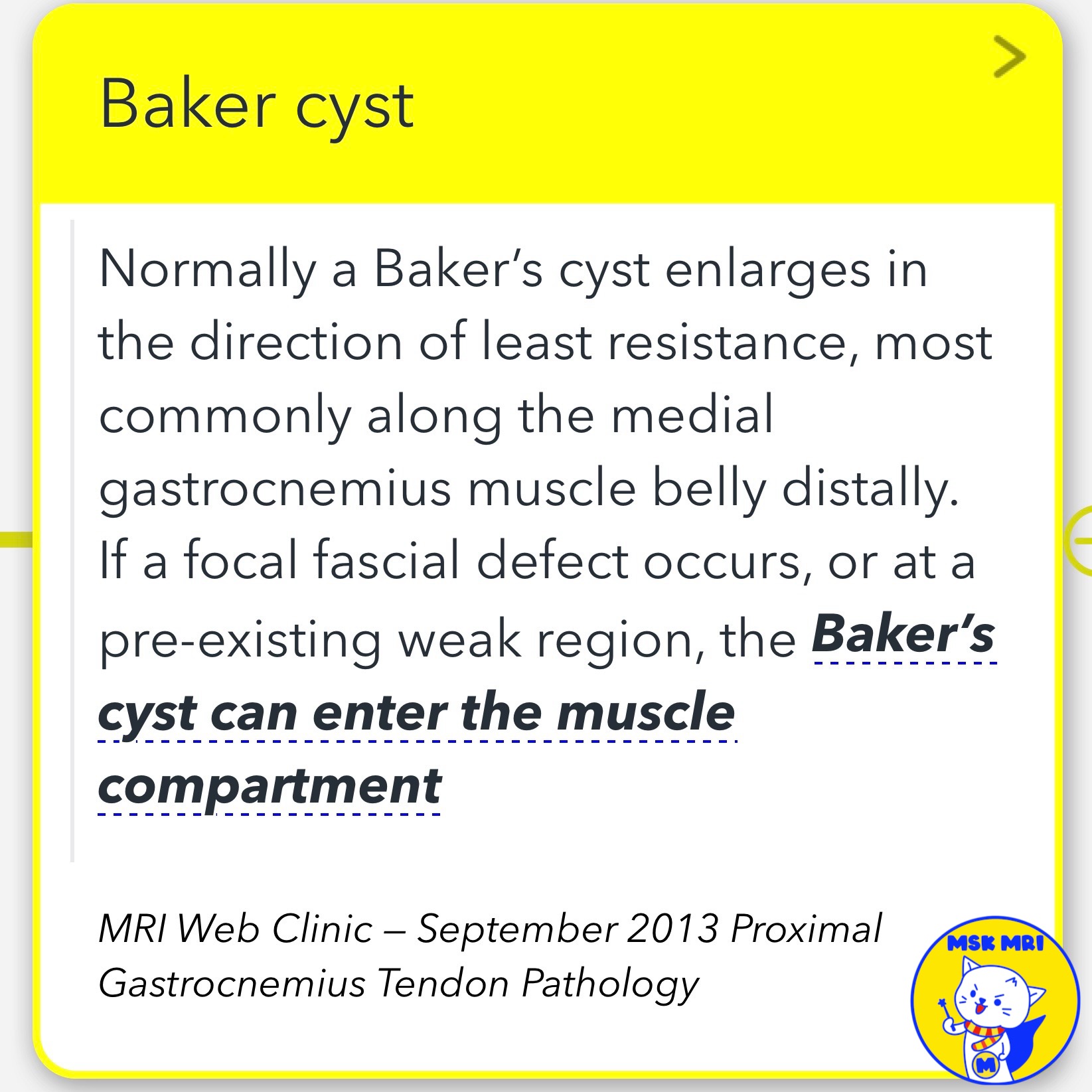
- Normally enlarges along medial gastrocnemius muscle distally
- Can enter muscle compartment if fascial defect or weak region present
- Dissecting cyst may present as tender fullness below knee joint
- Rare instance of cyst present both outside and within muscle compartment
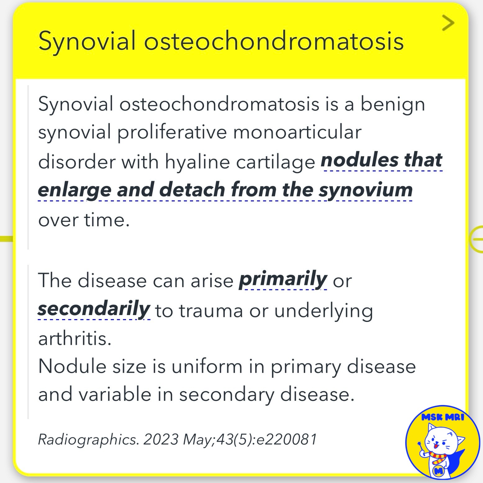
📌 Synovial Osteochondromatosis
- Benign synovial proliferative monoarticular disorder
- Hyaline cartilage nodules enlarge and detach from synovium over time
- Can be primary or secondary to trauma/arthritis
- Nodule size uniform in primary, variable in secondary disease
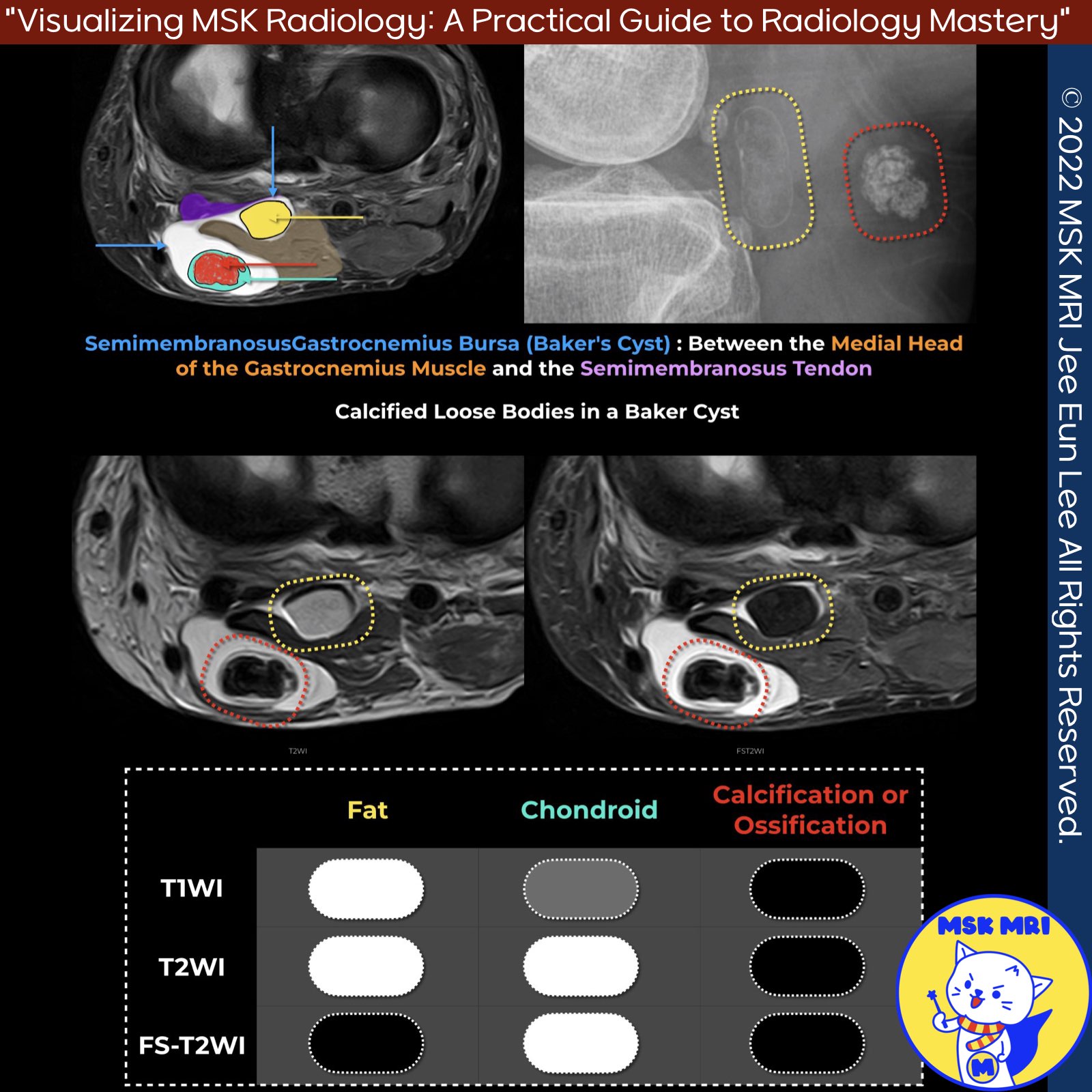
✅MRI signal depends on nodule composition:
- Chondroid: Isointense T1, high T2
- Calcified: Low T1, low T2
- Ossified with fatty marrow: High T1, high T2
Semin Musculoskelet Radiol. 2016 Feb;20(1):12-25
Radiographics. 2023 May;43(5):e220081
"Visualizing MSK Radiology: A Practical Guide to Radiology Mastery"
© 2022 MSK MRI Jee Eun Lee All Rights Reserved.
No unauthorized reproduction, redistribution, or use for AI training.
#BakersCyst, #DissectingBakersCyst, #SynovialOsteochondromatosis, #SynovialDisorders, #CartilageNodules, #SignalCharacteristics, #MonoarticularDisorder #MuscleFascialDefect,
'✅ Knee MRI Mastery > Chap 3.Collateral Ligaments' 카테고리의 다른 글
| (Fig 3-B.01) Three-Layer Approach to Lateral Knee (0) | 2024.05.19 |
|---|---|
| (Fig 3-A.53) MCL Bursitis ⎜Distinguishing from Grade I MCL Injury (0) | 2024.05.15 |
| (Fig 3-A.51) Semimembranosus Bursitis (0) | 2024.05.15 |
| (Fig 3-A.50) Pes Anserine Bursitis (0) | 2024.05.14 |
| (Fig 3-A.48) Osteomeniscal Impingement (0) | 2024.05.14 |
