Click the link to purchase on Amazon 🎉📚
==============================================
🎥 Check Out All Videos at Once! 📺
👉 Visit Visualizing MSK Blog to explore a wide range of videos! 🩻
https://visualizingmsk.blogspot.com/?view=magazine
📚 You can also find them on MSK MRI Blog and Naver Blog! 📖
https://www.instagram.com/msk_mri/
Click now to stay updated with the latest content! 🔍✨
==============================================
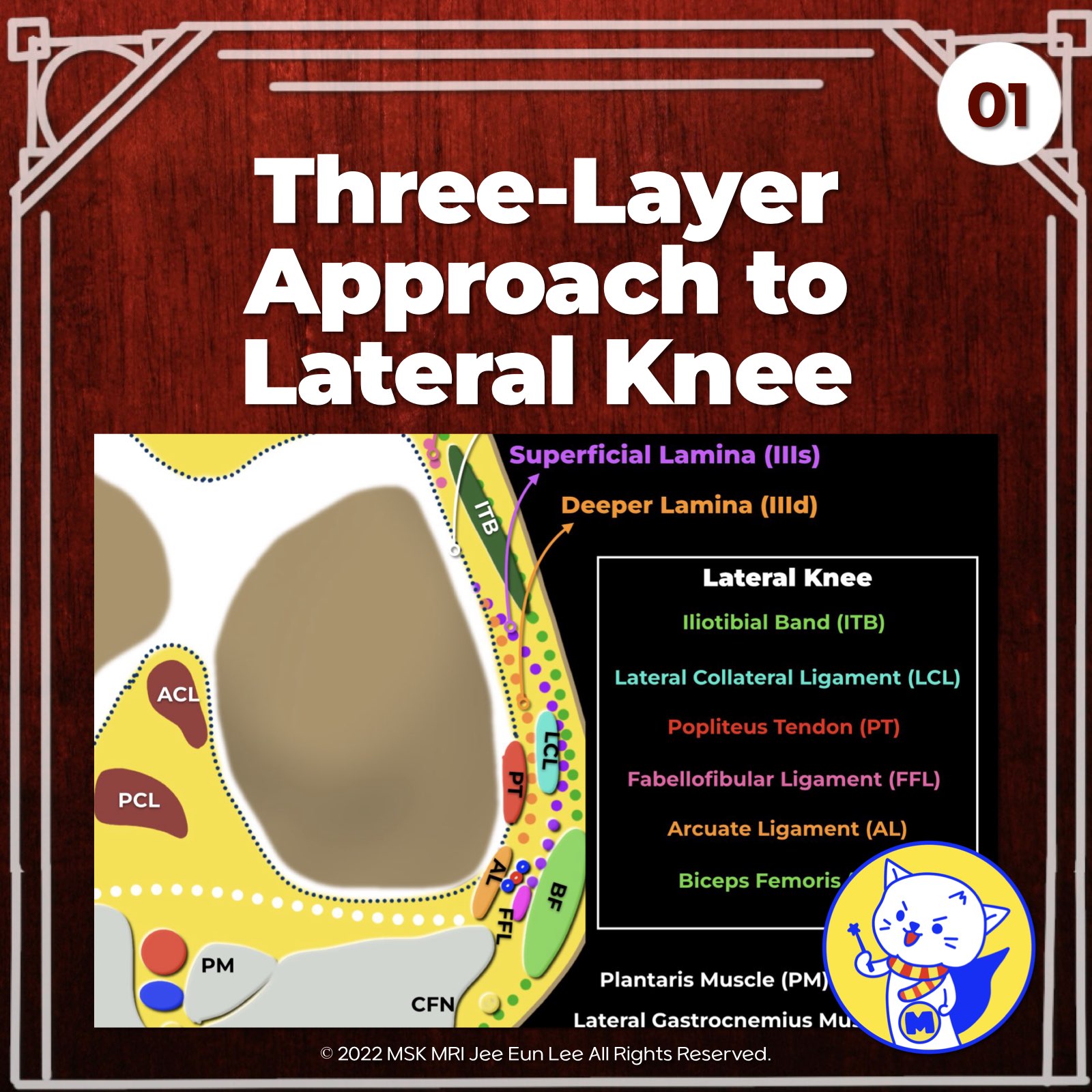
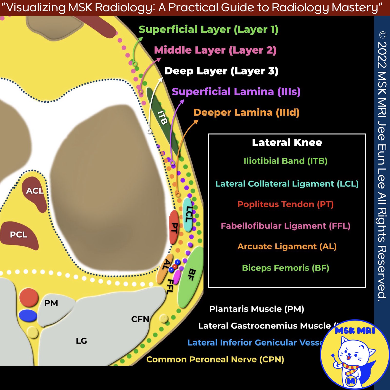
📌 The Posterolateral Corner of the Knee: Three-Layer Approach
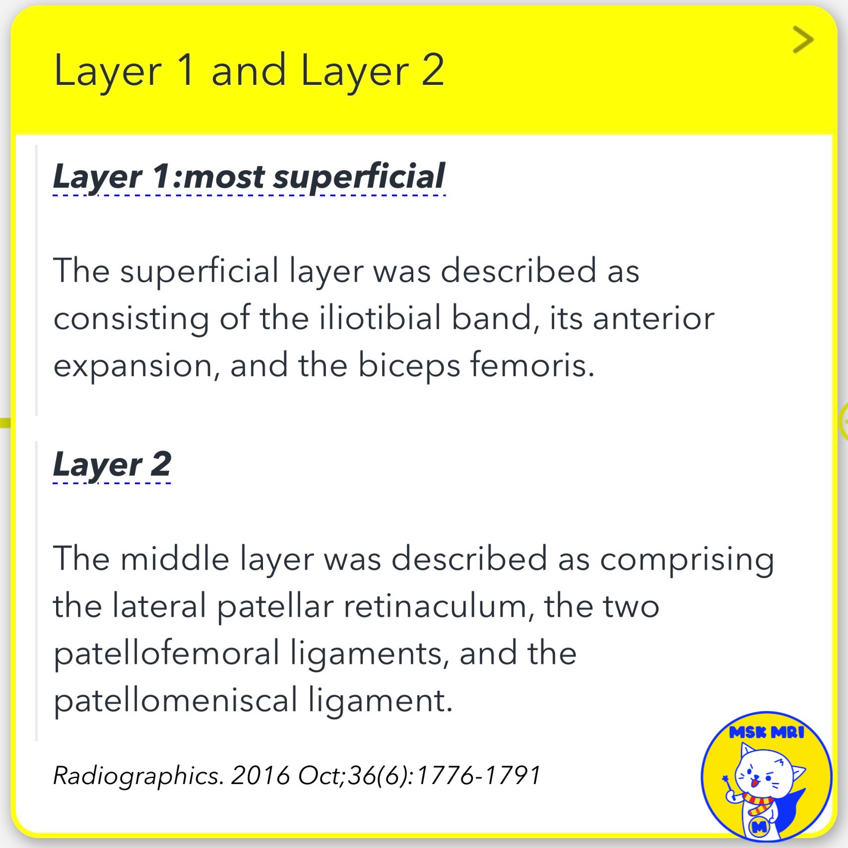
1️⃣ Superficial Layer (First Layer)
- Lateral fascia
- Iliotibial band
- Biceps femoris tendon
2️⃣ Middle Layer (Second Layer)
- Patellar retinaculum
- Patellofemoral ligament
- Patellomeniscal ligament
3️⃣ Deep Layer (Third Layer)
- Lateral collateral ligament (fibular collateral ligament)
- Lateral coronary ligament (lateral meniscotibial ligament)
- Arcuate ligament
- Popliteus tendon-muscle unit
- Popliteofibular ligament
- Fabellofibular ligament
- Lateral joint capsule with attachment to lateral meniscus edge
Note: The deep layer is the most anatomically variable and constitutes the posterolateral corner.
★ 1. Superficial Lamina - Travels superficial to lateral collateral ligament - Ends posteriorly at fabellofibular ligament
★ 2. Deep Lamina - Travels deep to lateral collateral ligament - Attaches to lateral meniscus edge, forming coronary ligament - Reaches arcuate ligament
Radiol Clin N Am 51 (2013) 413–432
"Visualizing MSK Radiology: A Practical Guide to Radiology Mastery"
© 2022 MSK MRI Jee Eun Lee All Rights Reserved.
No unauthorized reproduction, redistribution, or use for AI training.
#kneeanatomy, #posterolateralcorner, #lateralcollateralligament, #lateralcoronaryligament, #arcuateligament, #popliteustendon, #popliteofibularligament, #fabellofibularligament, #lateraljointcapsule, #lateralmeniscusattachment, #kneeanatomy, #posterolateralcornerstructures


'✅ Knee MRI Mastery > Chap 3.Collateral Ligaments' 카테고리의 다른 글
| (Fig 3-B.05) Lateral Collateral Ligament Anatomy: Part 1 (0) | 2024.05.20 |
|---|---|
| (Fig 3-B.02) Posterolateral Capsular Support Structures (0) | 2024.05.20 |
| (Fig 3-A.53) MCL Bursitis ⎜Distinguishing from Grade I MCL Injury (0) | 2024.05.15 |
| (Fig 3-A.52) Semimembranosus-Gastrocnemius Bursa: Baker Cyst, Synovial Osteochondromatosis (0) | 2024.05.15 |
| (Fig 3-A.51) Semimembranosus Bursitis (0) | 2024.05.15 |