Click the link to purchase on Amazon 🎉📚
==============================================
🎥 Check Out All Videos at Once! 📺
👉 Visit Visualizing MSK Blog to explore a wide range of videos! 🩻
https://visualizingmsk.blogspot.com/?view=magazine
📚 You can also find them on MSK MRI Blog and Naver Blog! 📖
https://www.instagram.com/msk_mri/
Click now to stay updated with the latest content! 🔍✨
==============================================
📌Anatomy of the Lateral Collateral Ligament (LCL) and Related Structures
1️⃣ Origin of the LCL
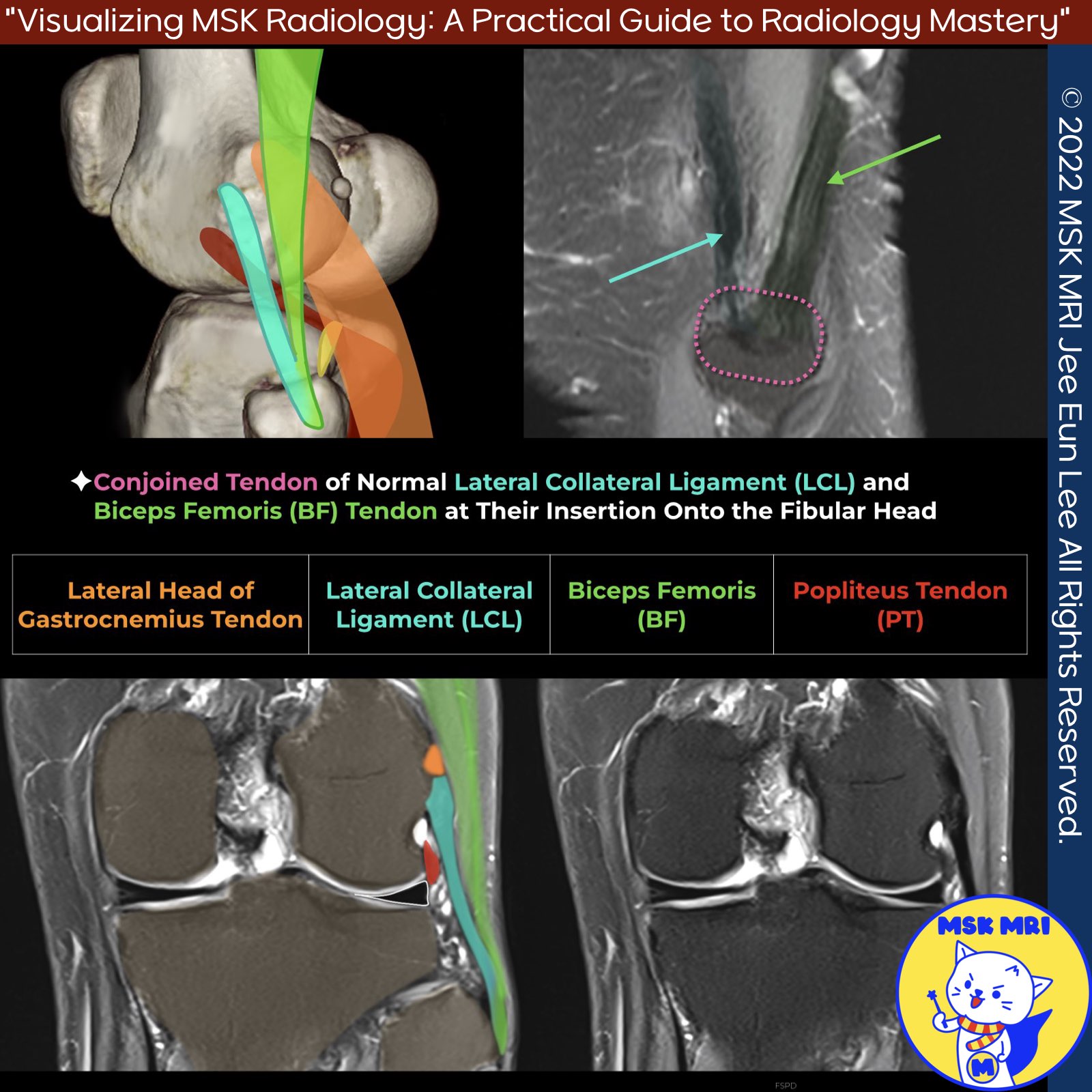
- The lateral collateral ligament (LCL) originates from the lateral side of the distal femur in a fanlike fashion, attaching between the lateral epicondyle and supracondylar process, just proximal and posterior to the lateral epicondyle, and anterior to the lateral head of the gastrocnemius muscle.
2️⃣ Insertion of the LCL
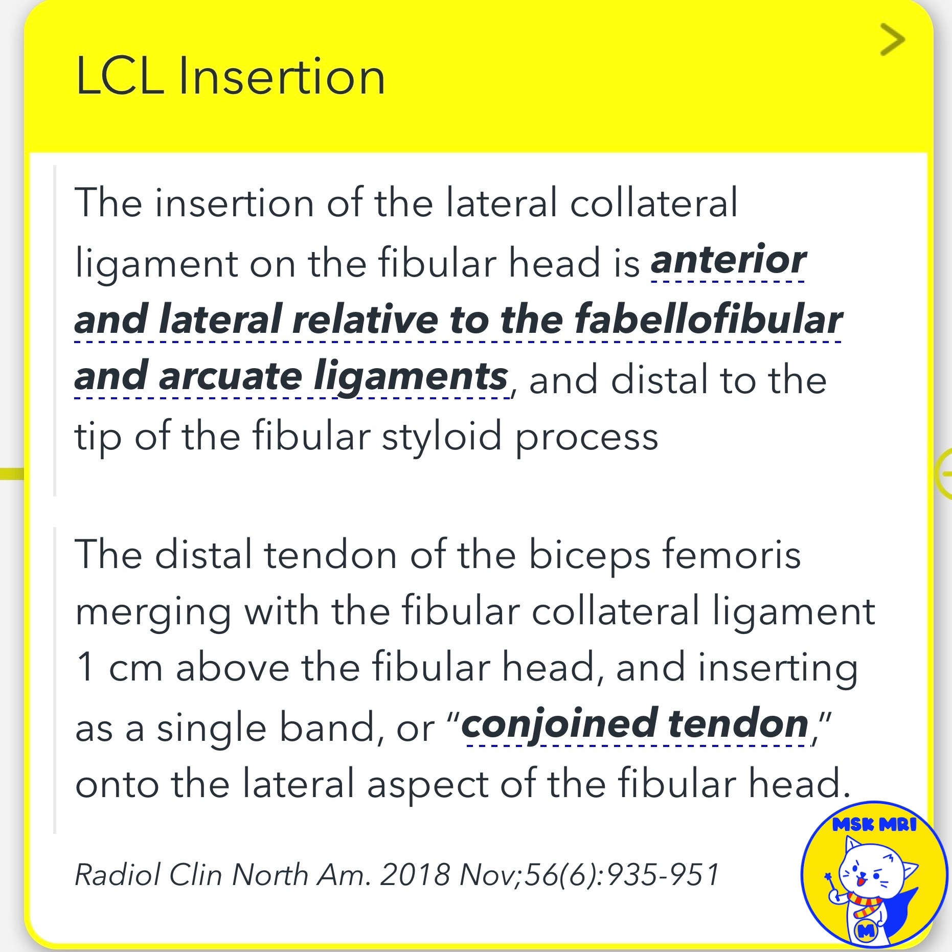
- The LCL inserts on the lateral aspect of the fibular head, anterior and lateral to the fabellofibular and arcuate ligament attachments, and distal to the fibular styloid process tip.
3️⃣ Lateral Gastrocnemius Tendon
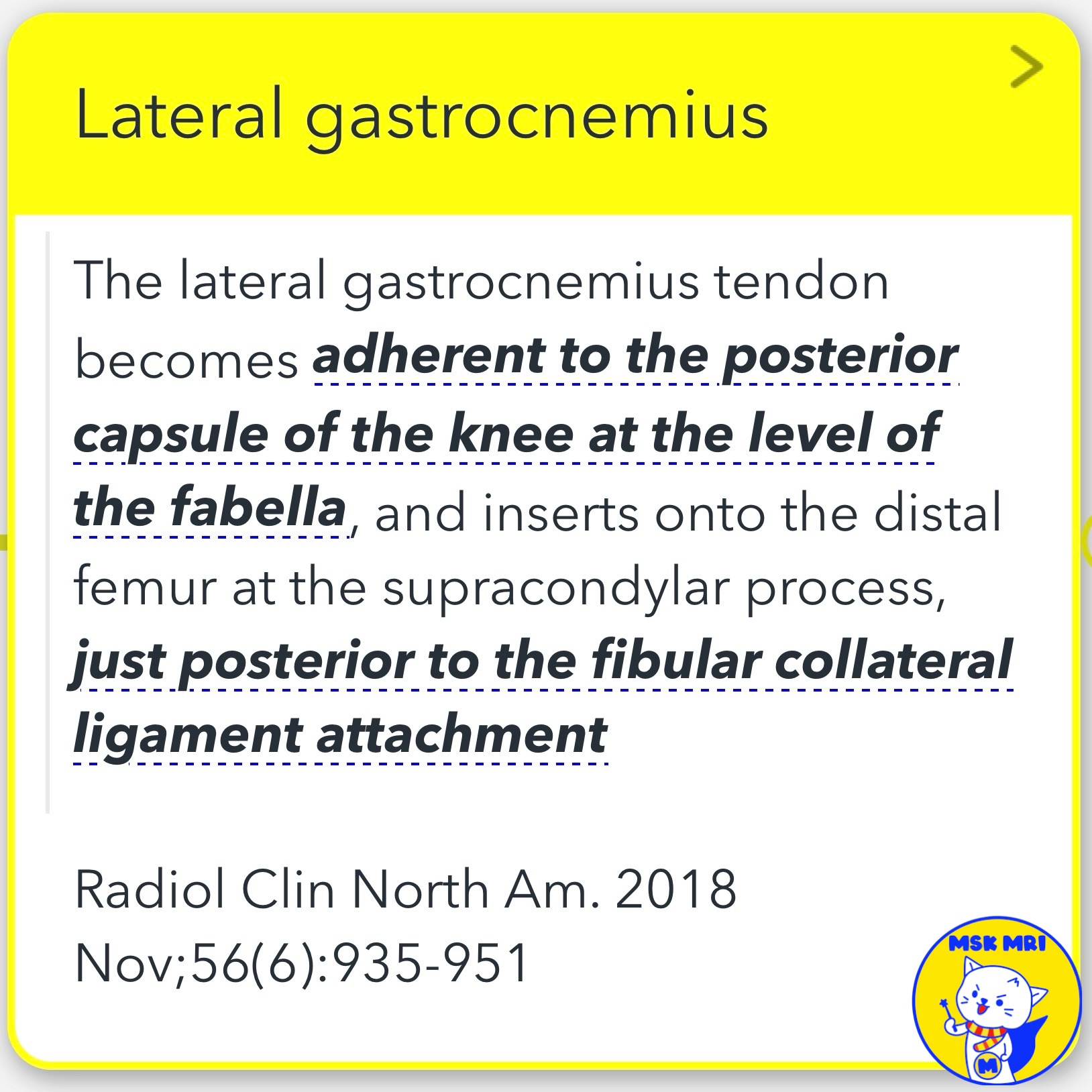
- The lateral gastrocnemius tendon becomes adherent to the posterior knee capsule at the fabella level, inserting on the distal femur at the supracondylar process, just posterior to the LCL femoral attachment.
4️⃣ Biceps Femoris
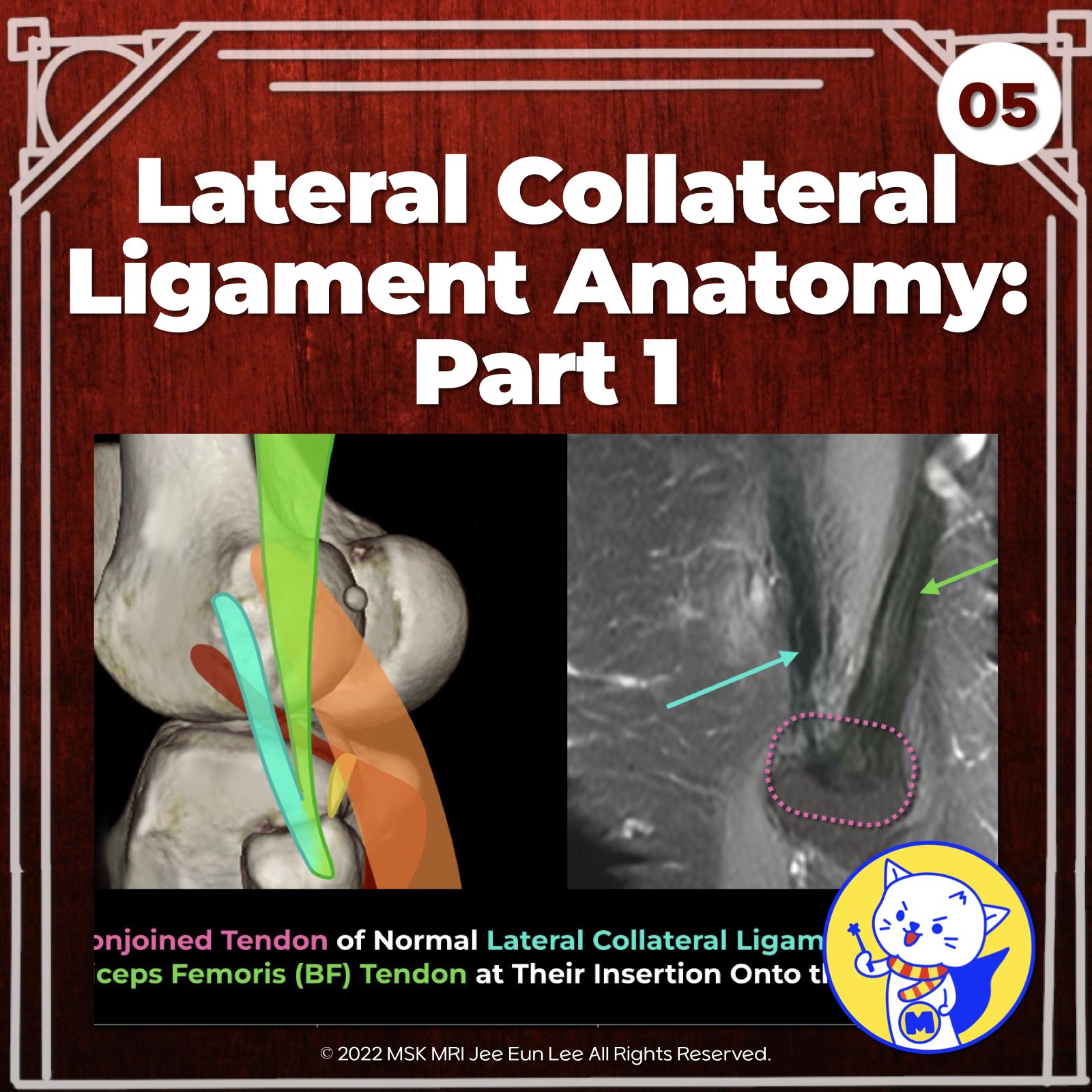
- Tendon The LCL and biceps femoris tendon blend into a conjoined tendon distally, inserting onto the fibular head.
5️⃣ Popliteus Tendon
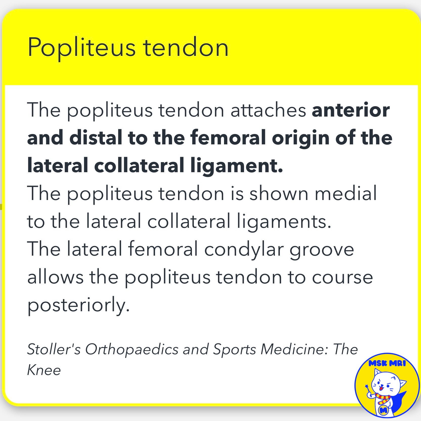
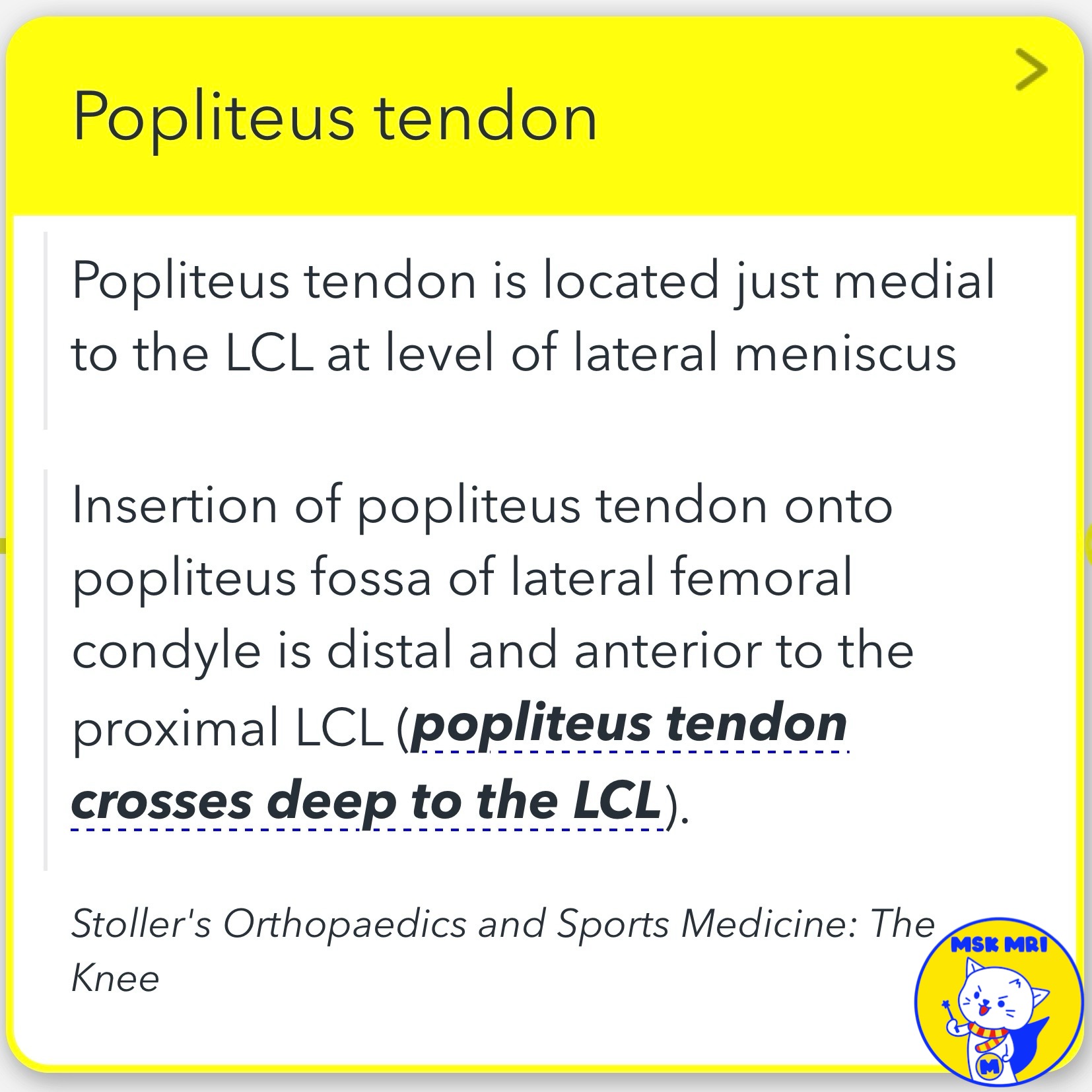
- The popliteus tendon attaches to the lateral femoral condyle anterior and distal to the LCL femoral origin.
- On coronal images, the popliteus insertion is seen below the lateral gastrocnemius and LCL origins.
- The lateral femoral condylar groove allows its posterior course.
Radiographics. 2016 Oct;36(6):1776-1791.
Radiol Clin North Am. 2018 Nov;56(6):935-951
Radiol Clin N Am 51 (2013) 413–432
Stoller's Orthopaedics and Sports Medicine: The Knee
RadioGraphics 2014; 34:496–513
"Visualizing MSK Radiology: A Practical Guide to Radiology Mastery"
© 2022 MSK MRI Jee Eun Lee All Rights Reserved.
No unauthorized reproduction, redistribution, or use for AI training.
#LateralCollateralLigament, #KneeAnatomy, #OrthopaedicSurgery, #LCLInjury, #LateralKneeInstability, #PopliteusTendon, #LateralGastrocnemiusTendon, #BicepsFemorisTendon, #FemoralLateralCondyle, #FibulaHead
'✅ Knee MRI Mastery > Chap 3.Collateral Ligaments' 카테고리의 다른 글
| (Fig 3-B.07) Mild Proximal LCL Injury (0) | 2024.05.20 |
|---|---|
| (Fig 3-B.06) Lateral Collateral Ligament Anatomy: Part 2 (0) | 2024.05.20 |
| (Fig 3-B.02) Posterolateral Capsular Support Structures (0) | 2024.05.20 |
| (Fig 3-B.01) Three-Layer Approach to Lateral Knee (0) | 2024.05.19 |
| (Fig 3-A.53) MCL Bursitis ⎜Distinguishing from Grade I MCL Injury (0) | 2024.05.15 |
