==============================================
⬇️✨⬇️🎉⬇️🔥⬇️📚⬇️
Click the link to purchase on Amazon 🎉📚
==============================================
🎥 Check Out All Videos at Once! 📺
👉 Visit Visualizing MSK Blog to explore a wide range of videos! 🩻
https://visualizingmsk.blogspot.com/?view=magazine
📚 You can also find them on MSK MRI Blog and Naver Blog! 📖
https://www.instagram.com/msk_mri/
Click now to stay updated with the latest content! 🔍✨
==============================================
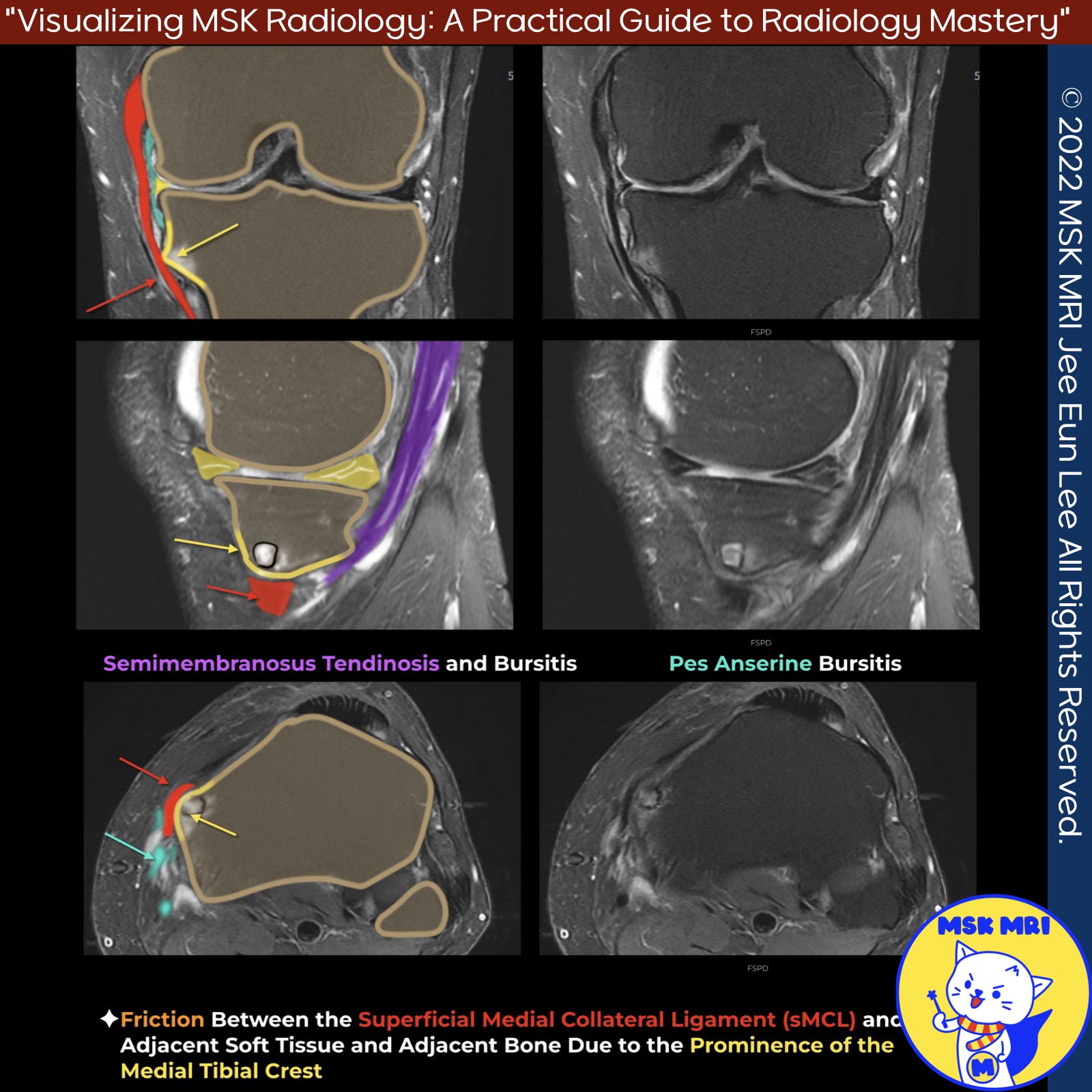
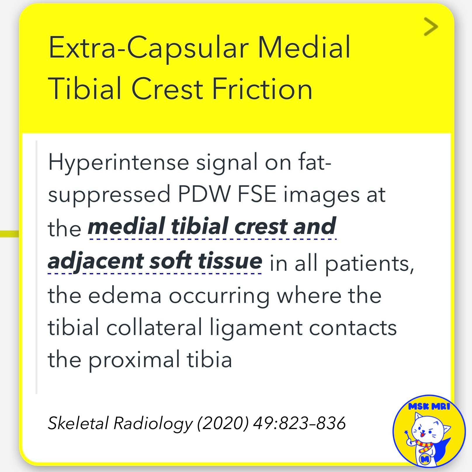
📌 Extra-Capsular Medial Tibial Crest Friction
✅ Clinical Presentation
- The condition is predominantly seen in young adults with a high level of physical activity, particularly athletes.
- Patients typically complain of pain and tenderness approximately 2.5 cm below the medial joint line.
✅ Imaging Findings
- It is suggested that prominence of the medial tibial crest due to a reduced medial tibial crest angle results in friction between the MCL and the adjacent bone.
- Hyperintense signal on fat-suppressed proton density-weighted fast spin-echo (PDW FSE) images at the medial tibial crest and adjacent soft tissue is observed in all patients, indicating edema occurring where the TCL contacts the proximal tibia.
✅Differential Diagnosis
- The only disorder that could probably mimic medial tibial crest friction syndrome with pain at this specific site could be pes anserine bursitis.
- However, pes anserine bursitis is located approximately 5 cm below the mid-articular line, whereas patients with medial tibial crest friction syndrome report pain 2.5 cm inferior to the knee joint space, as confirmed by clinical examination on-site, just prior to MRI examination.
- Moreover, pes anserine bursitis is typically found in obese females aged more than 50 years old.
Skeletal Radiology (2020) 49:823–836
Eur J Radiol. 2013 Nov;82(11):e703-6
"Visualizing MSK Radiology: A Practical Guide to Radiology Mastery"
© 2022 MSK MRI Jee Eun Lee All Rights Reserved.
No unauthorized reproduction, redistribution, or use for AI training.
#MedialTibialCrestFriction, #MCLFriction, #ExtraCapsularFriction, #SportsMedicine, #AthletesInjury, #TibialCrestAnatomy


'✅ Knee MRI Mastery > Chap 3.Collateral Ligaments' 카테고리의 다른 글
| (Fig 3-A.50) Pes Anserine Bursitis (0) | 2024.05.14 |
|---|---|
| (Fig 3-A.48) Osteomeniscal Impingement (0) | 2024.05.14 |
| (Fig 3-A.46) Adhesive Capsulitis of the knee (0) | 2024.05.14 |
| (Fig 3-A.45) Posteromedial Knee Friction Syndrome (0) | 2024.05.14 |
| (Fig 3-A.44) O’Donoghue’s Pentad Lesion (0) | 2024.05.13 |