Click the link to purchase on Amazon 🎉📚
==============================================
🎥 Check Out All Videos at Once! 📺
👉 Visit Visualizing MSK Blog to explore a wide range of videos! 🩻
https://visualizingmsk.blogspot.com/?view=magazine
📚 You can also find them on MSK MRI Blog and Naver Blog! 📖
https://www.instagram.com/msk_mri/
Click now to stay updated with the latest content! 🔍✨
==============================================
출처: https://mskmri.tistory.com/1056 [MSK MRI:티스토리]
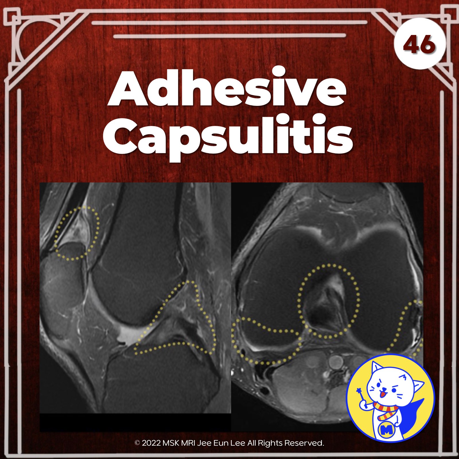
📌Adhesive Capsulitis
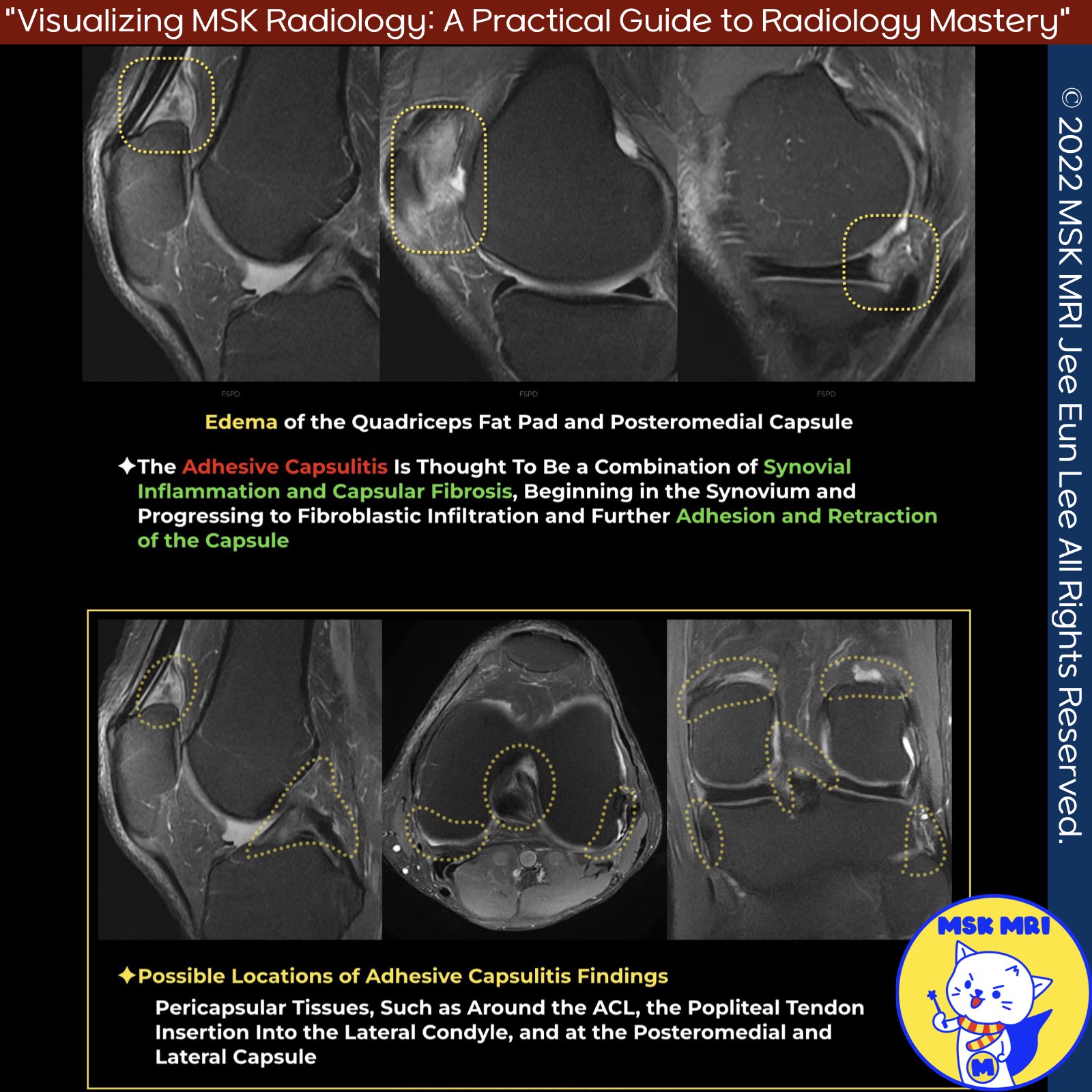
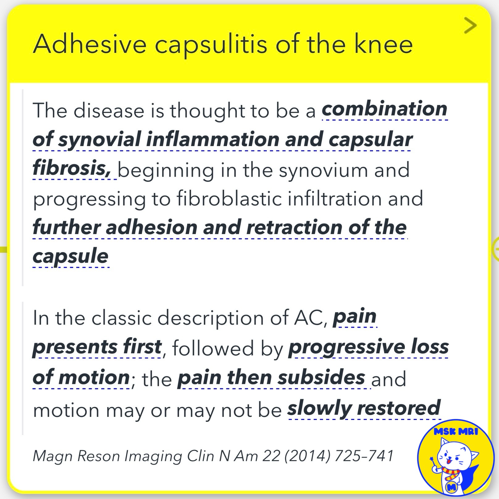
- Adhesive capsulitis (AC) is a condition characterized by synovial inflammation and capsular fibrosis, leading to fibroblastic infiltration, adhesion, and retraction of the capsule.
- It was traditionally associated with the glenohumeral (shoulder) joint, but recent evidence suggests it can affect other joints as well.
✅ Multi-Joint Involvement
- AC has been reported in joints such as the wrist, hip, and knee, indicating that it is not unique to the shoulder joint.
- Clinical Presentation: In the classic description of AC, pain presents first, followed by progressive loss of motion. The pain may subside, and motion may or may not be slowly restored.
✅ Imaging Findings:
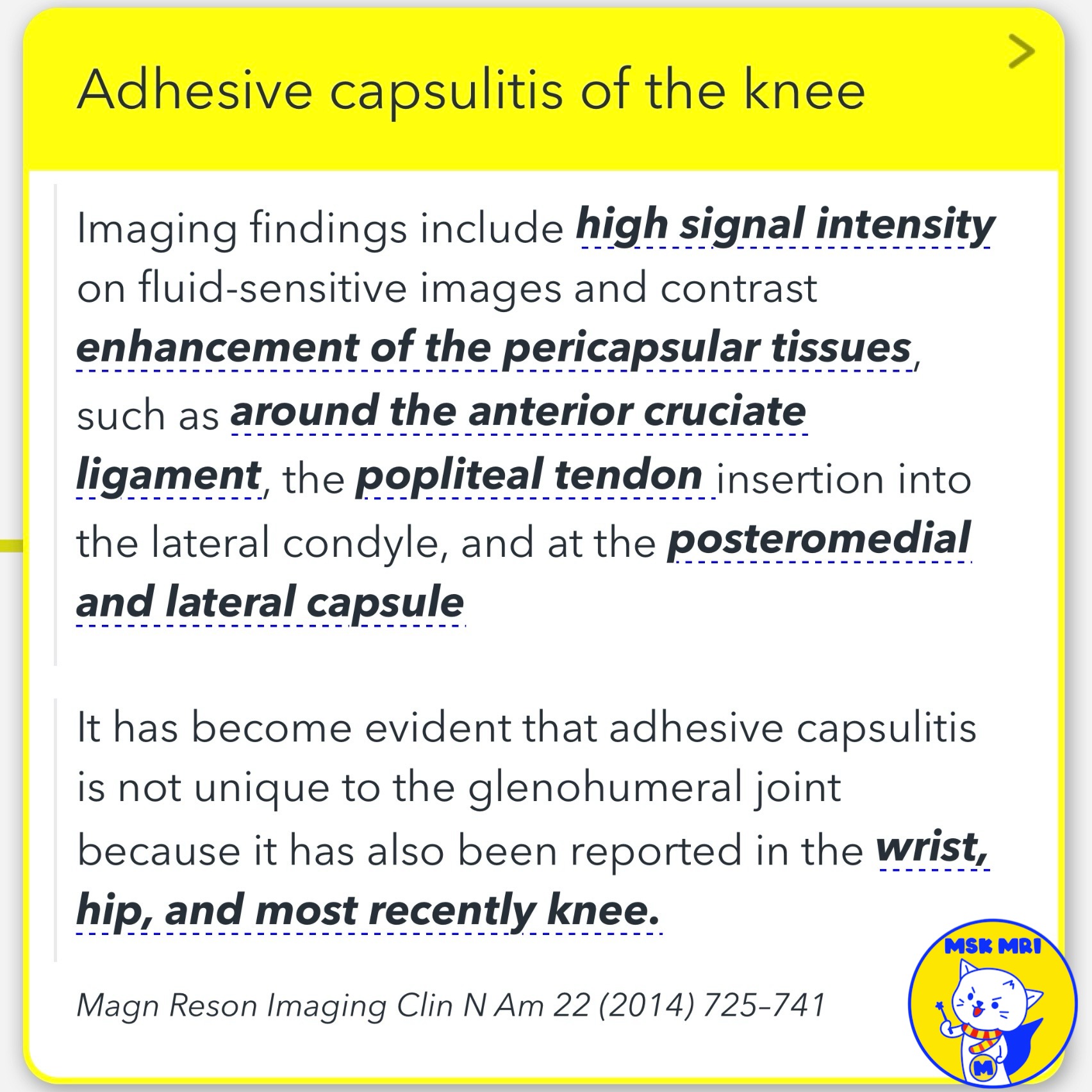
- Imaging findings in affected joints include high signal intensity on fluid-sensitive images and contrast enhancement of the pericapsular tissues, such as around the anterior cruciate ligament (ACL), popliteal tendon insertion, and the posteromedial and lateral capsule in the knee.
- De Abreu et al. reported a case of knee pain with high T2 signal diffusely along the ACL, suprapatellar fat pad, and posteromedial capsule. Fluorodeoxyglucose positron emission tomography/computed tomography revealed extensive enhancement of the same structures, as well as the posterior cruciate ligament (PCL), popliteus insertion, posterior capsule, and semimembranosus (SM) insertion.
✅ Diagnosis Confirmation:
- The diagnosis of AC in this case was confirmed or strengthened by biopsy, with no other findings in the affected knee and a normal contralateral knee.
Magn Reson Imaging Clin N Am 22 (2014) 725–741
Semin Musculoskelet Radiol. 2016 Feb;20(1):12-25
"Visualizing MSK Radiology: A Practical Guide to Radiology Mastery"
© 2022 MSK MRI Jee Eun Lee All Rights Reserved.
No unauthorized reproduction, redistribution, or use for AI training.
#AdhesiveCapsulitis, #CapsularFibrosis, #SynovialInflammation,#PericapsularTissues, #KneePain, #Fatpad,
'✅ Knee MRI Mastery > Chap 3.Collateral Ligaments' 카테고리의 다른 글
| (Fig 3-A.48) Osteomeniscal Impingement (0) | 2024.05.14 |
|---|---|
| (Fig 3-A.47) Extra-Capsular Medial Tibial Crest Friction (0) | 2024.05.14 |
| (Fig 3-A.45) Posteromedial Knee Friction Syndrome (0) | 2024.05.14 |
| (Fig 3-A.44) O’Donoghue’s Pentad Lesion (0) | 2024.05.13 |
| (Fig 3-A.43) O'Donoghue’s Triad Lesion (0) | 2024.05.13 |
