Click the link to purchase on Amazon 🎉📚
==============================================
🎥 Check Out All Videos at Once! 📺
👉 Visit Visualizing MSK Blog to explore a wide range of videos! 🩻
https://visualizingmsk.blogspot.com/?view=magazine
📚 You can also find them on MSK MRI Blog and Naver Blog! 📖
https://www.instagram.com/msk_mri/
Click now to stay updated with the latest content! 🔍✨
==============================================
📌 Popliteus Injuries
- Most popliteus tears are extra-articular, involving the muscle or myotendinous portion
- Less common: injuries to the tendon within the popliteal hiatus or near femoral insertion
- Musculotendinous junction or femoral insertion injuries are common in high-grade posterolateral corner (PLC) injuries
- Isolated popliteus injuries represent <10% of cases
✅ Imaging Findings Extra-articular tears:
- Muscle or myotendinous portion tears
- Strain/grade II lesion at myotendinous junction (white arrows)
- Extensive circumferential soft tissue edema
- Popliteus tendon appears intact
✅ MRI Findings Injury appearance varies by location and severity:
- Abnormal signal in popliteus muscle
- Irregular tendon contour with peritendinous edema
- Tendon avulsion from femoral attachment
Radiographics.2000 Oct;20 Spec No:S91-S102.
Radiol Clin North Am. 2018 Nov;56(6):935-951
"Visualizing MSK Radiology: A Practical Guide to Radiology Mastery"
© 2022 MSK MRI Jee Eun Lee All Rights Reserved.
No unauthorized reproduction, redistribution, or use for AI training.
#popliteusinjury, #popliteustear, #posterolateralcornerinjury, #plcinjury, #kneeinjury, #sportsinjury, #mri, #musculoskeletalmri,


'✅ Knee MRI Mastery > Chap 3.Collateral Ligaments' 카테고리의 다른 글
| (Fig 3-B.15) Popliteus Tendon Avulsion Fracture (0) | 2024.05.21 |
|---|---|
| (Fig 3-B.14) Intra-Articular Partial Tear of the Popliteus (0) | 2024.05.21 |
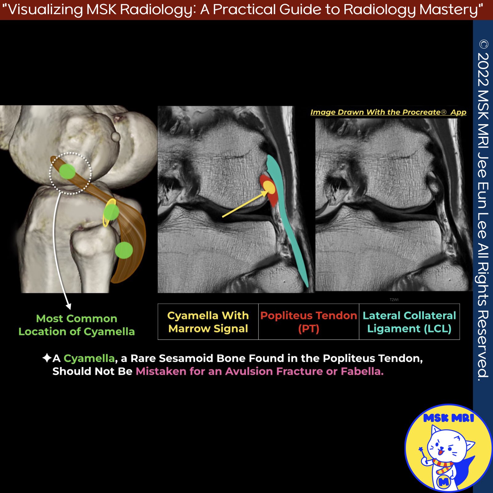
| (Fig 3-B.12) Cyamella vs Fabella (0) | 2024.05.21 |
| (Fig 3-B.11) Popliteus Musculotendinous Complex Anatomy (0) | 2024.05.21 |
| (Fig 3-B.10) Surrounding Popliteus Tendon Anatomy (0) | 2024.05.21 |