Click the link to purchase on Amazon 🎉📚
==============================================
🎥 Check Out All Videos at Once! 📺
👉 Visit Visualizing MSK Blog to explore a wide range of videos! 🩻
https://visualizingmsk.blogspot.com/?view=magazine
📚 You can also find them on MSK MRI Blog and Naver Blog! 📖
https://www.instagram.com/msk_mri/
Click now to stay updated with the latest content! 🔍✨
==============================================
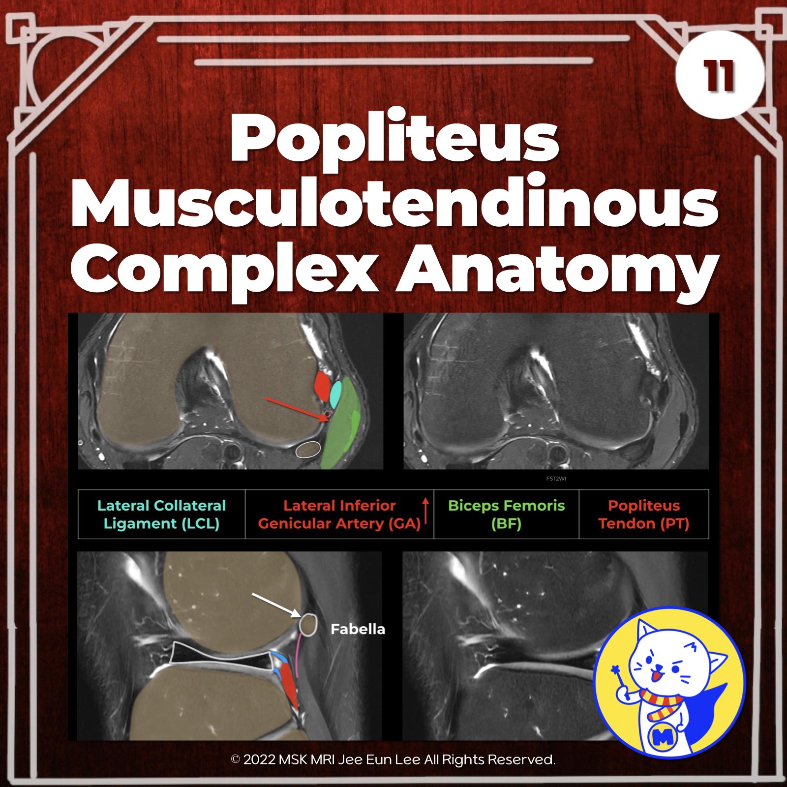
📌 Overview of Popliteus Musculotendinous Complex
The popliteus musculotendinous complex includes the popliteus tendon, muscle, and patellofemoral ligament. It provides resistance to external rotation during knee flexion and supports the posterior cruciate ligament by restraining posterior translation.
✅ Popliteus Tendon Pathway
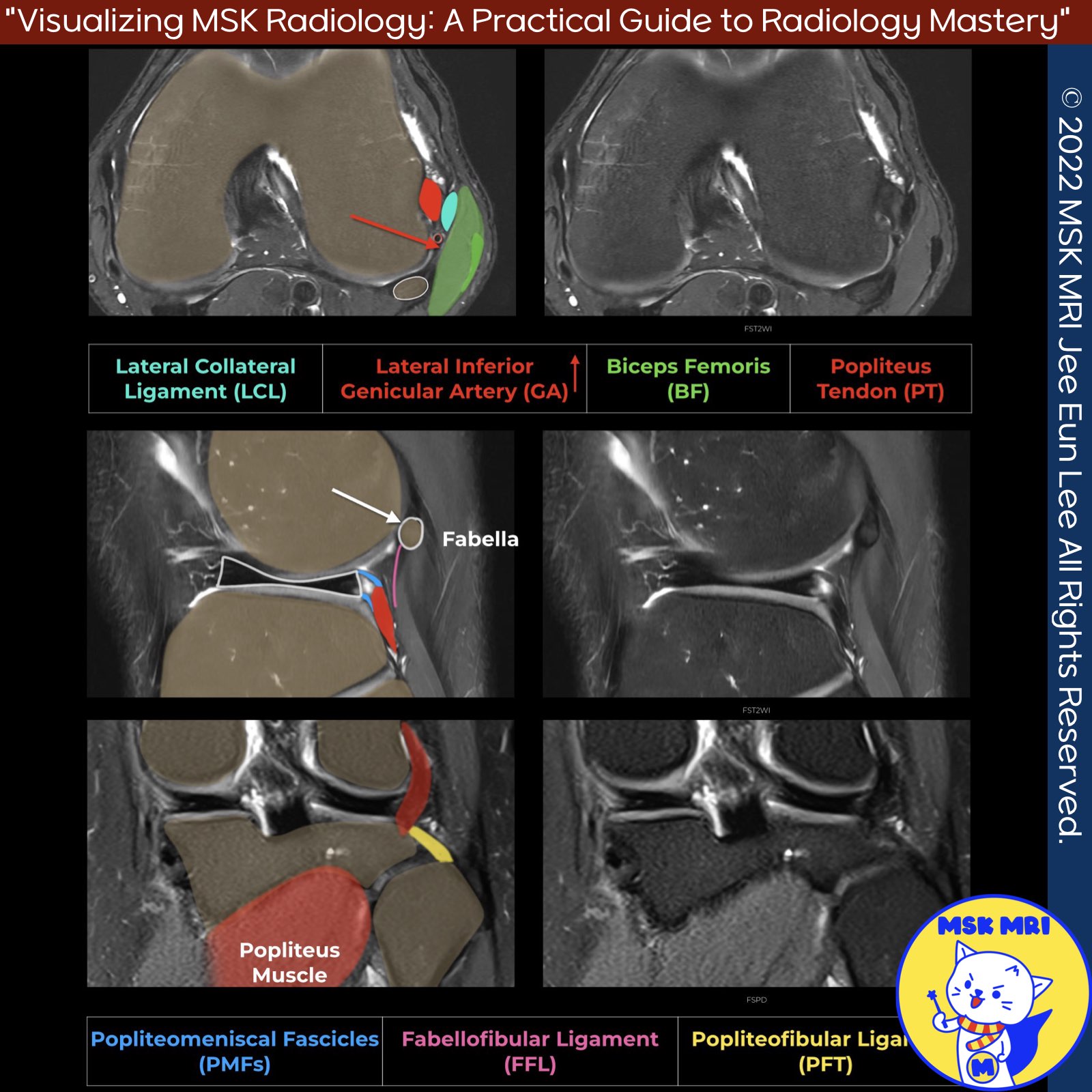
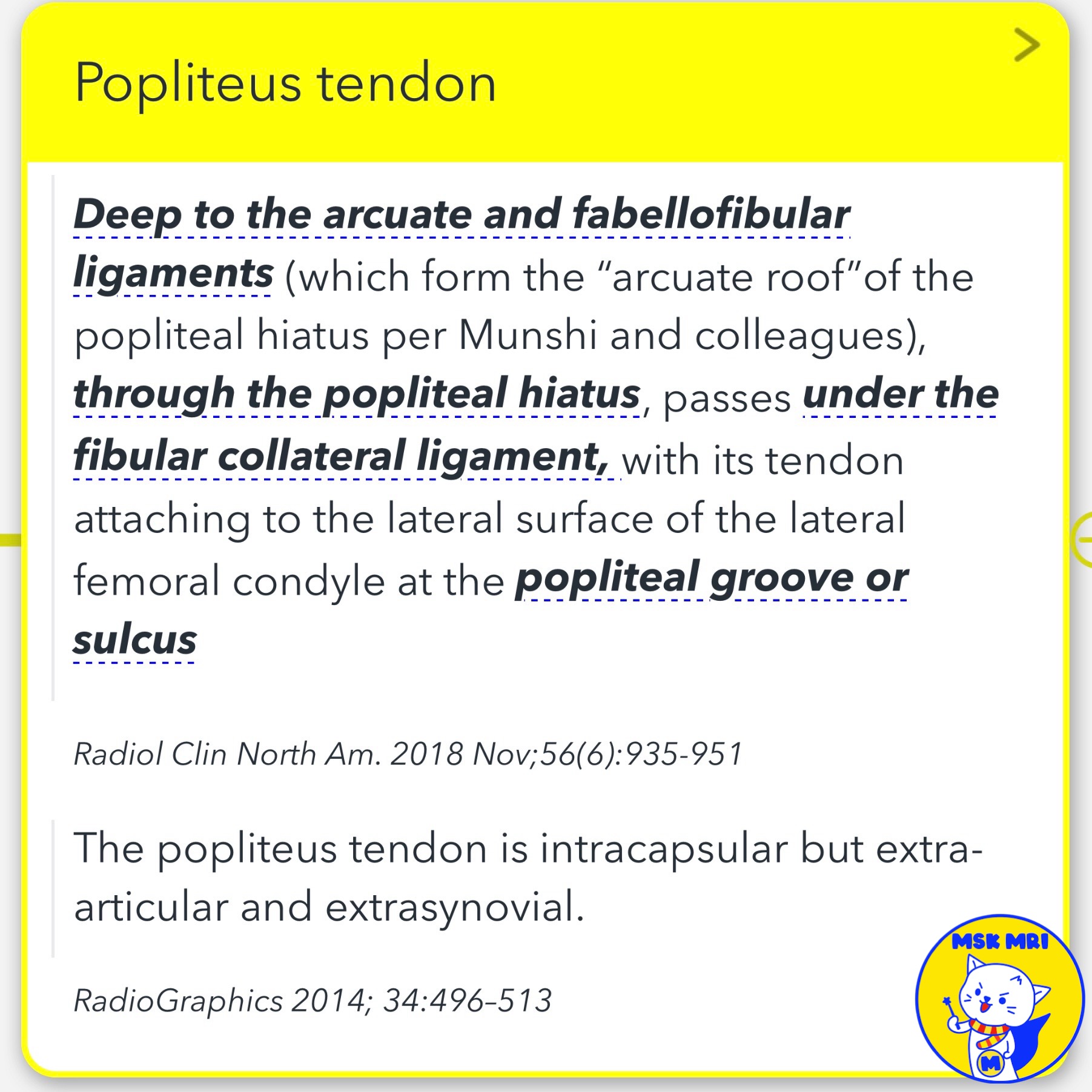
The popliteus tendon travels through the popliteal hiatus, bound by popliteomeniscal fascicles, and moves underneath the posterolateral joint capsule and arcuate ligament to become extracapsular.
✅ Popliteofibular Ligament
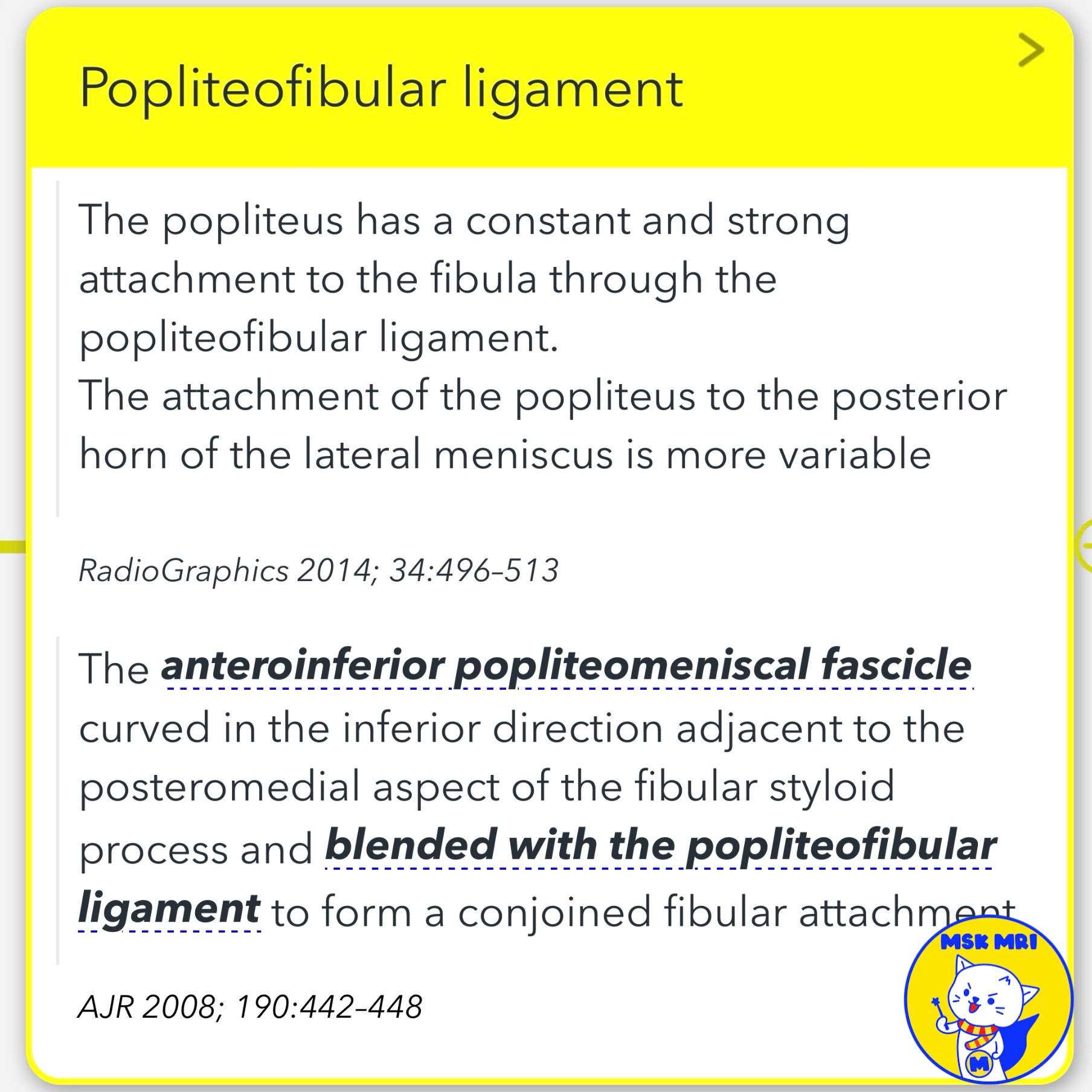
The popliteofibular ligament runs from the popliteus tendon near the myotendinous junction to the fibular styloid process, located posteromedial to the biceps femoris and LCL insertion. It can be identified by locating the lateral geniculate vessels on coronal images.
✅ Popliteomeniscal Fascicles
The popliteomeniscal fascicles are divided into three components:
- Anteroinferior (yellow): Forms the lateral floor of the popliteal hiatus, merging with the popliteofibular ligament.
- Posterosuperior (red): Forms the roof of the popliteal hiatus, attaching to the posterior joint capsule above the popliteus tendon.
- Posteroinferior (purple): A controversial structure.
✅ Popliteus muscle insertion
The popliteus tendon passes under the fibular collateral ligament and attaches to the lateral femoral condyle.
The popliteus muscle attaches to the fibula through the popliteofibular ligament and inserts on the posterior surface of the proximal tibia, just superior to the soleal line.
Radiographics. 2016 Oct;36(6):1776-1791
RadioGraphics 2014; 34:496–513
Radiol Clin North Am. 2018 Nov;56(6):935-951
Magn Reson Imaging Clin N Am 22 (2014) 493–516
AJR 2008; 190:442–448
"Visualizing MSK Radiology: A Practical Guide to Radiology Mastery"
© 2022 MSK MRI Jee Eun Lee All Rights Reserved.
No unauthorized reproduction, redistribution, or use for AI training.
#PopliteusComplex, #KneeFlexionMechanics, #PopliteusTendon, #PopliteofibularLigament, #PopliteomeniscalFascicles, #PosteriorCruciateSupport, #MusculotendinousAnatomy, #KneeStability, #OrthopedicAnatomy, #KneeLigaments
'✅ Knee MRI Mastery > Chap 3.Collateral Ligaments' 카테고리의 다른 글
| (Fig 3-B.13) Popliteus Myotendinous Junction Injury (0) | 2024.05.21 |
|---|---|
| (Fig 3-B.12) Cyamella vs Fabella (0) | 2024.05.21 |
| (Fig 3-B.10) Surrounding Popliteus Tendon Anatomy (0) | 2024.05.21 |
| (Fig 3-B.09) Complete Distal LCL Tear with Retraction (0) | 2024.05.21 |
| (Fig 3-B.08) Distal LCL Attachment Tear (0) | 2024.05.21 |


