Click the link to purchase on Amazon 🎉📚
==============================================
🎥 Check Out All Videos at Once! 📺
👉 Visit Visualizing MSK Blog to explore a wide range of videos! 🩻
https://visualizingmsk.blogspot.com/?view=magazine
📚 You can also find them on MSK MRI Blog and Naver Blog! 📖
https://www.instagram.com/msk_mri/
Click now to stay updated with the latest content! 🔍✨
==============================================
📌Posterolateral Corner (PLC) Injuries
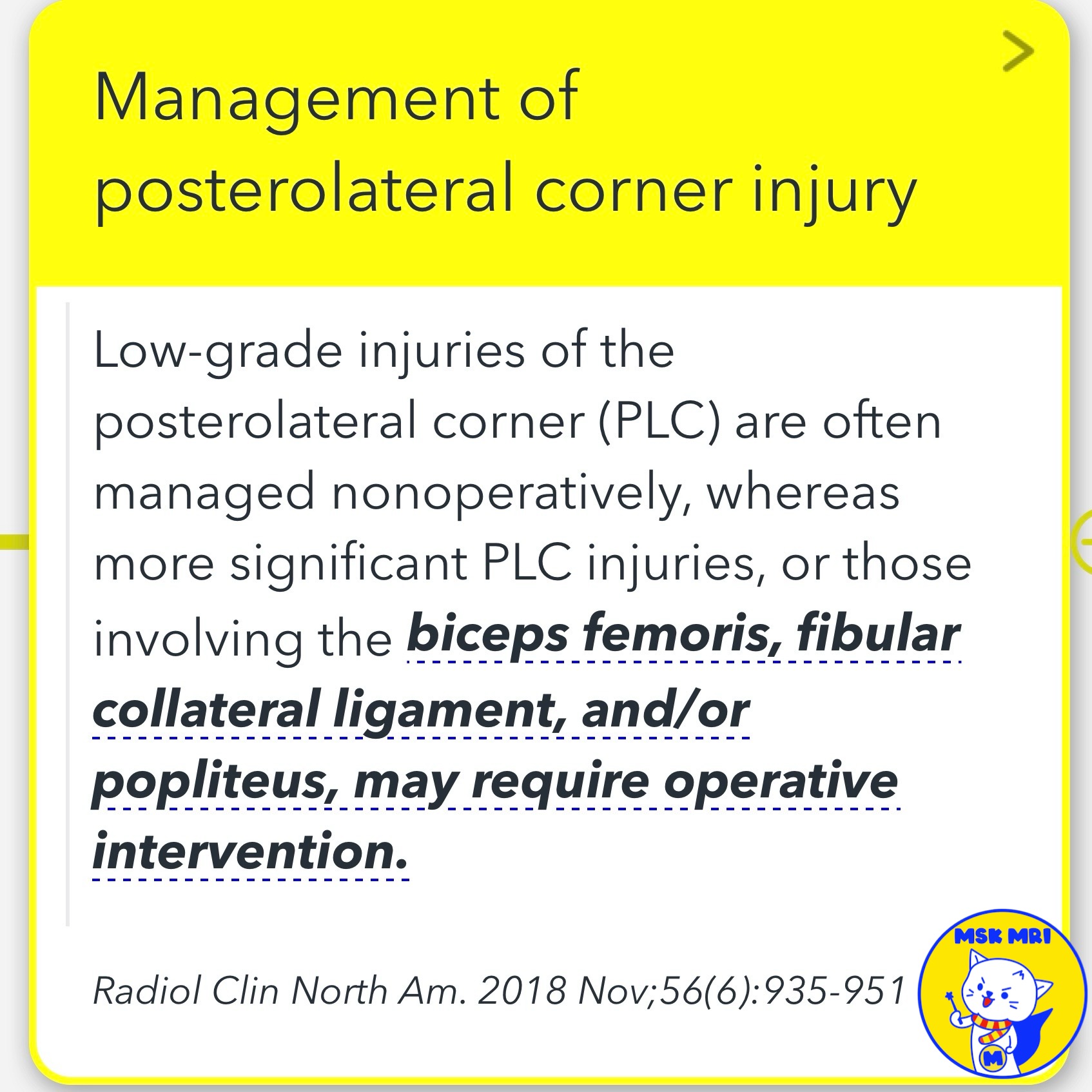
- Low-grade PLC injuries are often managed non-operatively
- Significant PLC injuries involving the biceps femoris, fibular collateral ligament, and/or popliteus may require surgical intervention
✅ MRI Assessment of PLC Injuries
- Evaluating PLC injuries with MRI is challenging due to anatomic complexity
- However, larger PLC structures are easy to assess with standard MRI techniques
- Direct visualization of small structure involvement on MRI is not necessary to report a clinically unstable PLC injury
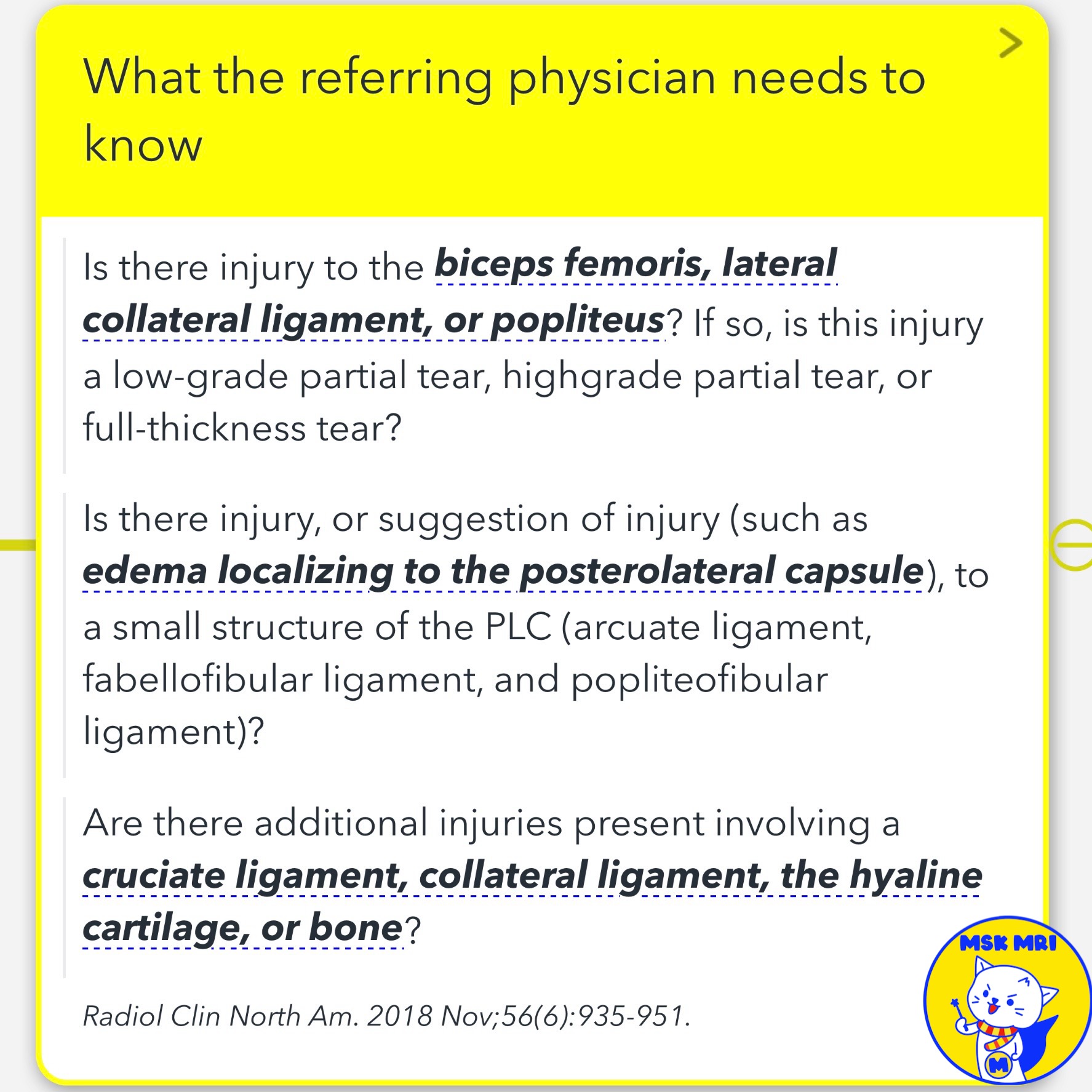
✅ Key Information Needed Extent of Injury
- Is there injury to the biceps femoris, lateral collateral ligament, or popliteus?
- If so, is it a low-grade partial tear, high-grade partial tear, or full-thickness tear?
- Is there injury or evidence of injury (e.g., edema) to smaller PLC structures like the arcuate ligament, fabellofibular ligament, or popliteofibular ligament?
- Are there additional injuries present involving a cruciate ligament, collateral ligament, hyaline cartilage, or bone?
Radiol Clin North Am. 2018 Nov;56(6):935-951.
"Visualizing MSK Radiology: A Practical Guide to Radiology Mastery"
© 2022 MSK MRI Jee Eun Lee All Rights Reserved.
No unauthorized reproduction, redistribution, or use for AI training.
#plcinjury, #posterolateralcornerinjury, #kneeanatomy, #kneesurgery, #orthopedics, #sportsmedicine, #mri, #bicepsfemoris, #fibularcollateralligament, #popliteus

'✅ Knee MRI Mastery > Chap 3.Collateral Ligaments' 카테고리의 다른 글
| (Fig 3-B.11) Popliteus Musculotendinous Complex Anatomy (0) | 2024.05.21 |
|---|---|
| (Fig 3-B.10) Surrounding Popliteus Tendon Anatomy (0) | 2024.05.21 |
| (Fig 3-B.08) Distal LCL Attachment Tear (0) | 2024.05.21 |
| (Fig 3-B.07) Mild Proximal LCL Injury (0) | 2024.05.20 |
| (Fig 3-B.06) Lateral Collateral Ligament Anatomy: Part 2 (0) | 2024.05.20 |