Click the link to purchase on Amazon 🎉📚
==============================================
🎥 Check Out All Videos at Once! 📺
👉 Visit Visualizing MSK Blog to explore a wide range of videos! 🩻
https://visualizingmsk.blogspot.com/?view=magazine
📚 You can also find them on MSK MRI Blog and Naver Blog! 📖
https://www.instagram.com/msk_mri/
Click now to stay updated with the latest content! 🔍✨
==============================================
📌MRI Findings in Ligament Injuries
✅ Classification of Ligament Injuries
- At MR imaging, ligament injuries can be classified as:
- These often correspond to the clinical grading of grade I, II, and III injuries, respectively.
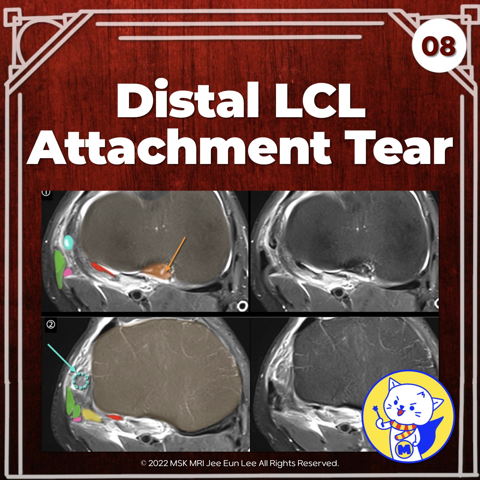
✅ Lateral Collateral Ligament (LCL) Injury Patterns
- Most common injury patterns of the fibular collateral ligament (LCL) include:
- According to Juhng et al., avulsion of the LCL usually occurs at its distal attachment from the head of the fibula, rather than its proximal femoral attachment.
- In complete LCL disruptions, the ligament demonstrates a wavy or serpiginous contour and loss of ligamentous continuity on MRI.
✅Associated Cruciate Ligament Injuries
- Isolated injuries to the posterolateral corner (PLC) of the knee are rare.
- PLC injuries usually occur in conjunction with injury to a cruciate ligament (ACL or PCL).
RadioGraphics 2016; 36:1776–1791
Radiol Clin North Am. 2018 Nov;56(6):935-951
Stoller's Orthopaedics and Sports Medicine: The Knee
"Visualizing MSK Radiology: A Practical Guide to Radiology Mastery"
© 2022 MSK MRI Jee Eun Lee All Rights Reserved.
No unauthorized reproduction, redistribution, or use for AI training.
#LCLTear, #PosterolateralCornerInjury, #PCLTear, #LigamentInjury, #CruciateLigamentInjury, #AvulsionFracture, #SoftTissueAvulsion, #WavyContourMRI, #DisruptedLigamentMRI, #KneeTrauma


'✅ Knee MRI Mastery > Chap 3.Collateral Ligaments' 카테고리의 다른 글
| (Fig 3-B.10) Surrounding Popliteus Tendon Anatomy (0) | 2024.05.21 |
|---|---|
| (Fig 3-B.09) Complete Distal LCL Tear with Retraction (0) | 2024.05.21 |
| (Fig 3-B.07) Mild Proximal LCL Injury (0) | 2024.05.20 |
| (Fig 3-B.06) Lateral Collateral Ligament Anatomy: Part 2 (0) | 2024.05.20 |
| (Fig 3-B.05) Lateral Collateral Ligament Anatomy: Part 1 (0) | 2024.05.20 |