Click the link to purchase on Amazon 🎉📚
==============================================
🎥 Check Out All Videos at Once! 📺
👉 Visit Visualizing MSK Blog to explore a wide range of videos! 🩻
https://visualizingmsk.blogspot.com/?view=magazine
📚 You can also find them on MSK MRI Blog and Naver Blog! 📖
https://www.instagram.com/msk_mri/
Click now to stay updated with the latest content! 🔍✨
==============================================
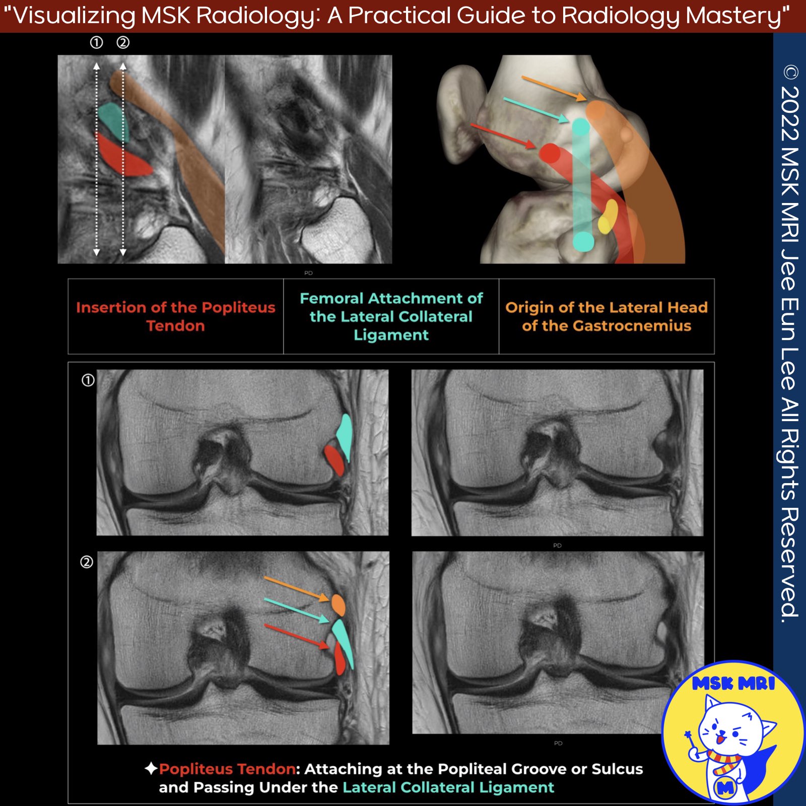
✅ Intra-Articular Popliteus Anatomy
- Knowing the intra-articular popliteus tendon anatomy is crucial.
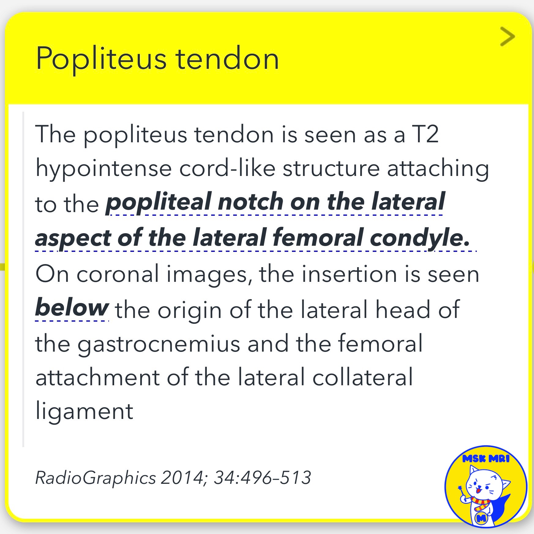
- The popliteus tendon attaches to the popliteus fossa of the lateral femoral condyle, anterior and distal to the femoral origin of the lateral collateral ligament (LCL). So the popliteus tendon is seen first, followed by the LCL.
- Moving further posteriorly, the lateral gastrocnemius tendon inserts onto the distal femur at the supracondylar process, just posterior to the LCL attachment.
✅ Popliteus Tendon Tears
- Most popliteus strains are extra-articular, involving the muscular or myotendinous portion.
- Popliteus tendon tears can be intra-articular at the level of the popliteal hiatus and at or near the femoral attachment.
- Intra-articular tears can be dealt with arthroscopically.
- The degree of tendon retraction in full-thickness tears is important.
RadioGraphics 2014; 34:496–513
"Visualizing MSK Radiology: A Practical Guide to Radiology Mastery"
© 2022 MSK MRI Jee Eun Lee All Rights Reserved.
No unauthorized reproduction, redistribution, or use for AI training.
#LateralCollateralLigament, #KneeAnatomy, #PopliteusTendon, #LateralGastrocnemiusTendon, #FemoralLateralCondyle, #popliteustendon, #lateralcollateralligament, #gastrocnemiustendon, #popliteusfossa, #femoralcondyle, #tendoninjuries,


'✅ Knee MRI Mastery > Chap 3.Collateral Ligaments' 카테고리의 다른 글
| (Fig 3-B.12) Cyamella vs Fabella (0) | 2024.05.21 |
|---|---|
| (Fig 3-B.11) Popliteus Musculotendinous Complex Anatomy (0) | 2024.05.21 |
| (Fig 3-B.09) Complete Distal LCL Tear with Retraction (0) | 2024.05.21 |
| (Fig 3-B.08) Distal LCL Attachment Tear (0) | 2024.05.21 |
| (Fig 3-B.07) Mild Proximal LCL Injury (0) | 2024.05.20 |