Click the link to purchase on Amazon 🎉📚
==============================================
🎥 Check Out All Videos at Once! 📺
👉 Visit Visualizing MSK Blog to explore a wide range of videos! 🩻
https://visualizingmsk.blogspot.com/?view=magazine
📚 You can also find them on MSK MRI Blog and Naver Blog! 📖
https://www.instagram.com/msk_mri/
Click now to stay updated with the latest content! 🔍✨
==============================================
📌 Iliotibial Band Anatomy
- The iliotibial band (ITB or IT band) is a thick band of fascia along the lateral aspect of the thigh, representing a thickening of the fascia lata.
Distal Insertions
- At least 5 distal insertions have been described around the lateral knee, attaching to the distal femur, patella, proximal tibia, and joint capsule.
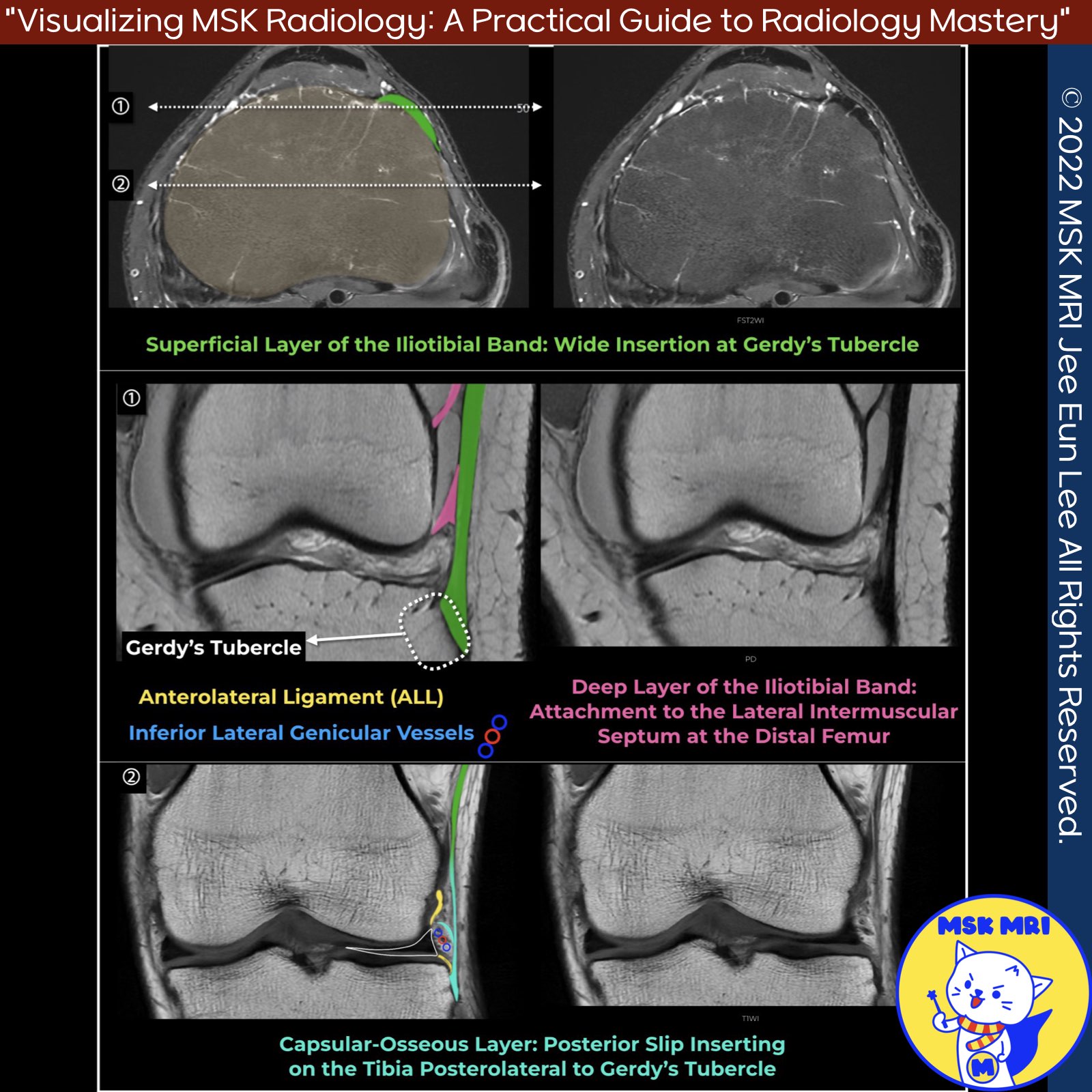
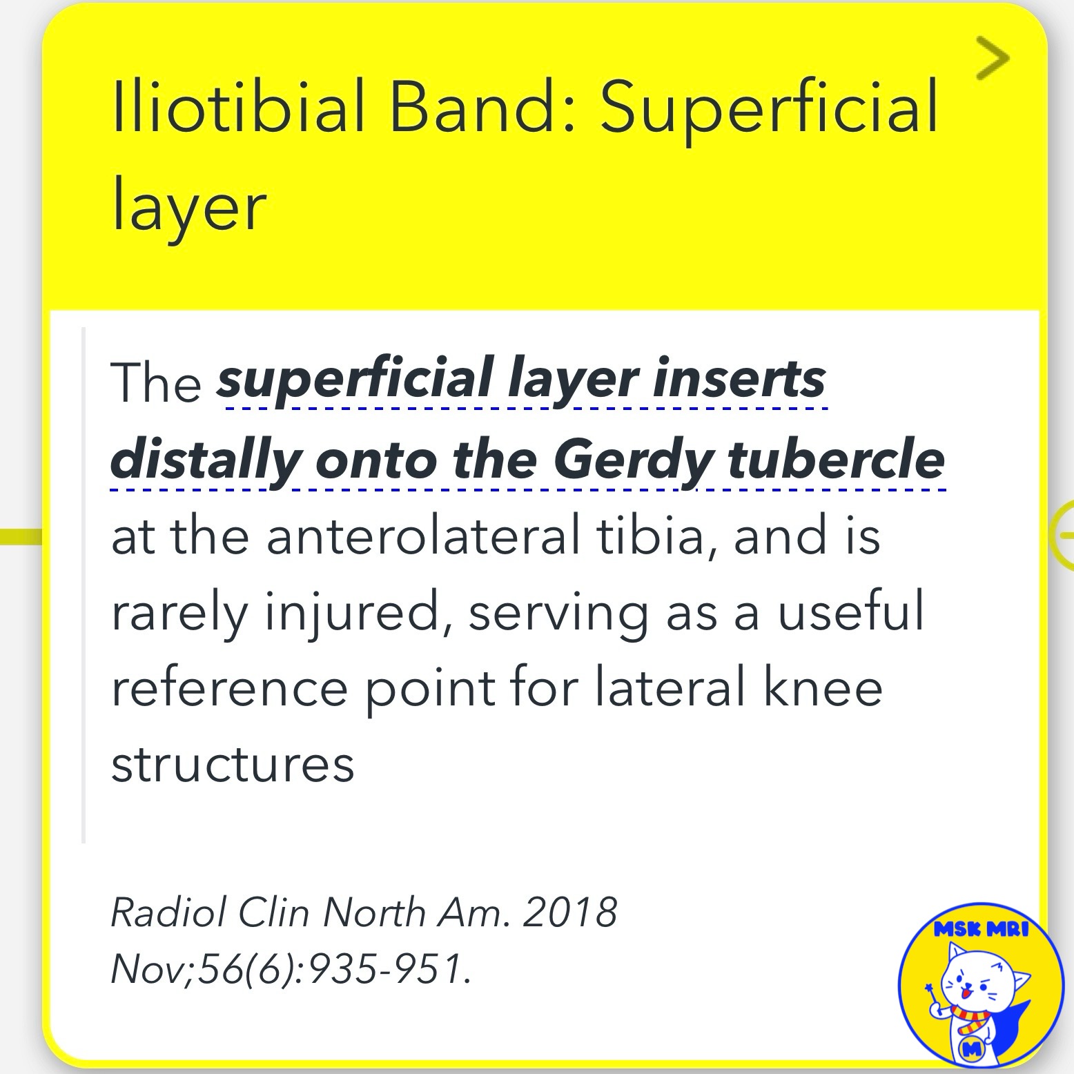
✅Superficial Layer
- Main tendinous component
- Inserts onto Gerdy's tubercle on anterior lateral tibia
✅Deep Layer
- Attaches superficial layer to lateral supracondylar tubercle of femur
- Blends into intermuscular septum at distal femur
✅Capsulo-Osseous Layer
- Situated deep and posterior to deep layer
- Forms a sling over lateral femoral condyle
- Inserts onto tibial tuberosity posterior and proximal to Gerdy's tubercle
- Some consider it the same as the anterolateral ligament
Radiol Clin North Am. 2018 Nov;56(6):935-951.
Skeletal Radiol. 2017 May;46(5):605-622
"Visualizing MSK Radiology: A Practical Guide to Radiology Mastery"
© 2022 MSK MRI Jee Eun Lee All Rights Reserved.
No unauthorized reproduction, redistribution, or use for AI training.


#ITBandAnatomy, #LateralKneeAnatomy, #OrthopedicAnatomy, #KneeInjuries, #RunningInjuries, #ITBFS, #LateralKneePain
'✅ Knee MRI Mastery > Chap 3.Collateral Ligaments' 카테고리의 다른 글
| (Fig 3-B.34) Iliotibial Band Friction Syndrome (0) | 2024.05.24 |
|---|---|
| (Fig 3-B.33) Iliotibial Band Injury from Acute Trauma (0) | 2024.05.23 |
| (Fig 3-B.31) Anterolateral rotary instability (0) | 2024.05.23 |
| (Fig 3-B.29) Segond Fracture (0) | 2024.05.23 |
| (Fig 3-B.28) Proximal Anterolateral Ligament Tear (0) | 2024.05.23 |