Click the link to purchase on Amazon 🎉📚
==============================================
🎥 Check Out All Videos at Once! 📺
👉 Visit Visualizing MSK Blog to explore a wide range of videos! 🩻
https://visualizingmsk.blogspot.com/?view=magazine
📚 You can also find them on MSK MRI Blog and Naver Blog! 📖
https://www.instagram.com/msk_mri/
Click now to stay updated with the latest content! 🔍✨
==============================================
✅ Patellar Insertion of the MPML and MPTL
The MPTL inserts into the proximal tibia and the distal pole of the patella, while the MPML inserts into the medial meniscus and the distal pole of the patella. These structures often have closely related or combined insertions in the patella (Arthroscopy. 2017 Oct;33(10):1862-1873). Additionally, the MPTL and MPML attach near the patellar tendon and patellar articular cartilage (Am J Sports Med. 2018 Jan;46(1):153-162).
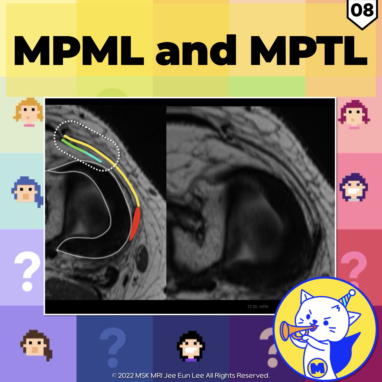
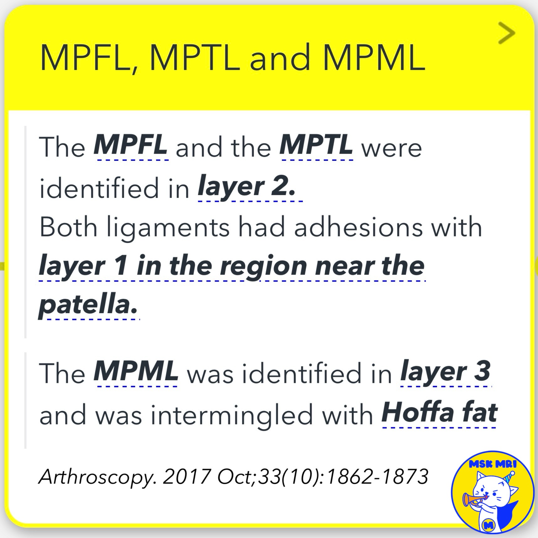
✅ Layer Identification and Relationships
The MPFL and MPTL are found in layer 2, with adhesions to layer 1 near the patella. The MPML is located in layer 3, intermingled with Hoffa's fat pad (Arthroscopy. 2017 Oct;33(10):1862-1873). The medial patellar retinaculum, described as having 2 or 3 layers, can be challenging to distinguish on MRI in normal patients (Magn Reson Imaging Clin N Am 22 (2014) 601–620).
✅ Distal Medial Patellar Restraints
The MPML and MPTL extend from the inferomedial patella to the medial meniscus and anteromedial tibia, respectively. Despite being considered secondary stabilizers, these ligaments significantly contribute to resisting lateral patellar translation, tilt, and rotation during knee flexion. MPFL reconstructions may now be combined with MPTL reconstruction for patients with patellar instability (Radiographics. 2023 Jun;43(6)
✅ Medial Patellomeniscal Ligament (MPML)
The MPML is located within layer III and is visible at the medial meniscal level as a hypointense taut band, continuous with the joint capsule. Its meniscal insertion is typically in the anterior horn or the transition from the anterior horn to the body (Skeletal Radiol (2012) 41:137–148; Arthroscopy. 2017 Oct;33(10):1862-1873).
✅ Medial Retinaculum and MPFL
The medial retinaculum is formed by the fusion of the superficial (layer 1) and intermediate (layer 2) layers of the medial knee supporting structures. The MPFL and medial retinaculum are often indistinguishable on imaging and are collectively described as the MPFL/medial retinacular complex or the MPFL as the most superior fibers of this complex (Skeletal Radiol. 2018 Aug;47(8):1069-1086).
"Visualizing MSK Radiology: A Practical Guide to Radiology Mastery"
© 2022 MSK MRI Jee Eun Lee All Rights Reserved.
No unauthorized reproduction, redistribution, or use for AI training.
#MedialRetinaculum, #MPFL, #MPML, #MPTL, #PatellarInsertion, #KneeLigaments, #KneeImaging, #OrthopedicRadiology, #PatellarStabilizers, #KneeAnatomy