Click the link to purchase on Amazon 🎉📚
==============================================
🎥 Check Out All Videos at Once! 📺
👉 Visit Visualizing MSK Blog to explore a wide range of videos! 🩻
https://visualizingmsk.blogspot.com/?view=magazine
📚 You can also find them on MSK MRI Blog and Naver Blog! 📖
https://www.instagram.com/msk_mri/
Click now to stay updated with the latest content! 🔍✨
==============================================
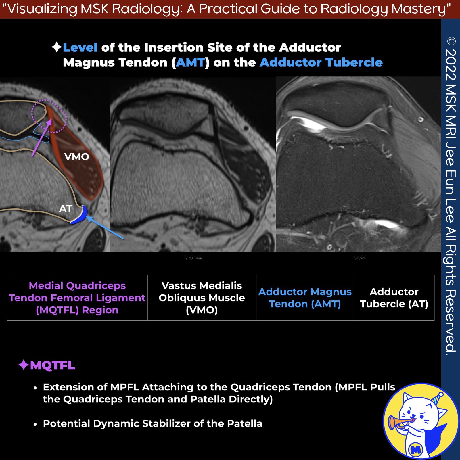
1️⃣ Medial Quadriceps Tendon Femoral Ligament
- The Medial Quadriceps Tendon Femoral Ligament (MQTFL) At the axial level of the insertion of the adductor magnus tendon (AMT) on the adductor tubercle, we can visualize the vastus medialis obliquus muscle, adductor magnus tendon, and adductor tubercle.
- Proximal to the MPFL and patella are oblique ligamentous fibers extending to insert into the distal quadriceps, termed the medial quadriceps tendon femoral ligament (MQTFL), outlined by the purple dotted line.
- Role of the MQTFL The MQTFL may contribute to patellar stability, particularly in full knee extension.
- Surgical Reconstruction
With modern surgical techniques, it is now possible to reconstruct the MPFL, the MQTFL, or both of these proximal medial patellar restraints.
Radiographics. 2023 Jun;43(6):e220177
Am J Sports Med. 2018 Jan;46(1):153-162
"Visualizing MSK Radiology: A Practical Guide to Radiology Mastery"
© 2022 MSK MRI Jee Eun Lee All Rights Reserved.
No unauthorized reproduction, redistribution, or use for AI training.
#mpflanatomy, #quadricepstendonfemoral, #patellofemoralcomplex, #patellarstability, #patellarmalalignment, #patellartracking, #mpflsurgery, #kneemri, #sportsmed, #orthoanatomy