Click the link to purchase on Amazon 🎉📚
==============================================
🎥 Check Out All Videos at Once! 📺
👉 Visit Visualizing MSK Blog to explore a wide range of videos! 🩻
https://visualizingmsk.blogspot.com/?view=magazine
📚 You can also find them on MSK MRI Blog and Naver Blog! 📖
https://www.instagram.com/msk_mri/
Click now to stay updated with the latest content! 🔍✨
==============================================
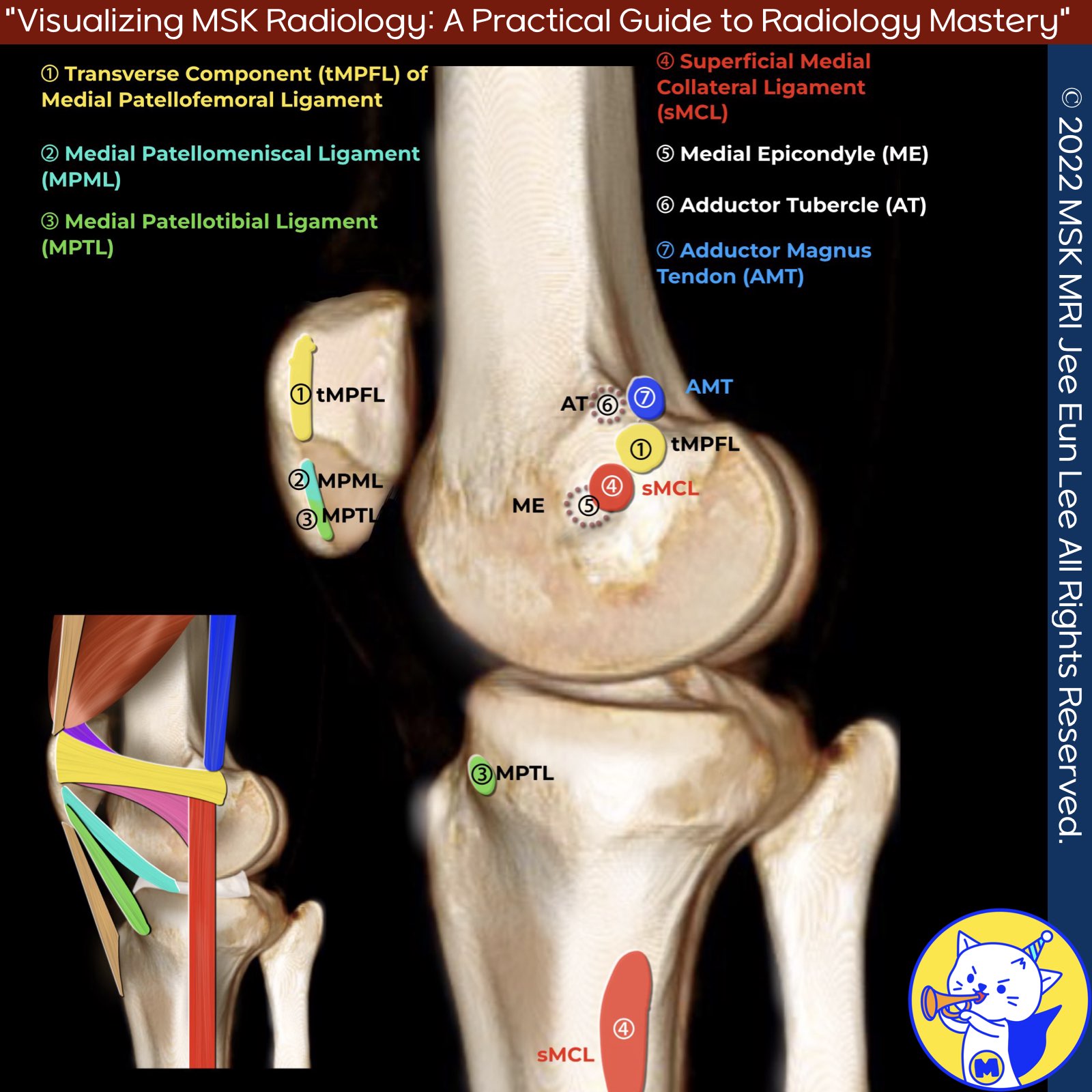
📌 Attachments of the Medial Patellofemoral Ligament Complex.
1️⃣ Transverse component of the MPFL (tMPFL)
- The femoral attachment of the tMPFL was assessed immediately below the level of the adductor tubercle (AT) and just above the superior attachment of the sMCL as described by LaPrade
- MPFL is generally described as a horizontally oriented, ribbonlike ligament that spans from the medial patella (proximal two-thirds) to the adjacent medial femur (most commonly near the physeal scar, at a sulcus between the medial femoral epicondyle and the adductor tubercle)
✅ Adductor tubercle
The adductor magnus tendon attached in an osseous depression an average of 3.0 mm (range, 1.8 to 4.6 mm) posterior and 2.7 mm (range, 1.6 to 4.3 mm) proximal to the adductor tubercle and did not attach directly to the adductor tubercle
2️⃣ Patellar insertion of the MPML and MPTL
- The MPTL inserts into the proximal tibia and into the distal pole of the patella.
- The MPML inserts into the medial meniscus and into the distal pole of the patella.
- The MPTL and the MPML joined to form a combined insertion in the patella in 5 knees. In 3 knees, the insertion of the MPML was proximal to the MPTL, and in 1 sample, the insertion of the MPTL was proximal to the MPML.
- The current study also revealed the close proximity of the MPTL and MPML attachment to the patellar tendon and patellar articular cartilage
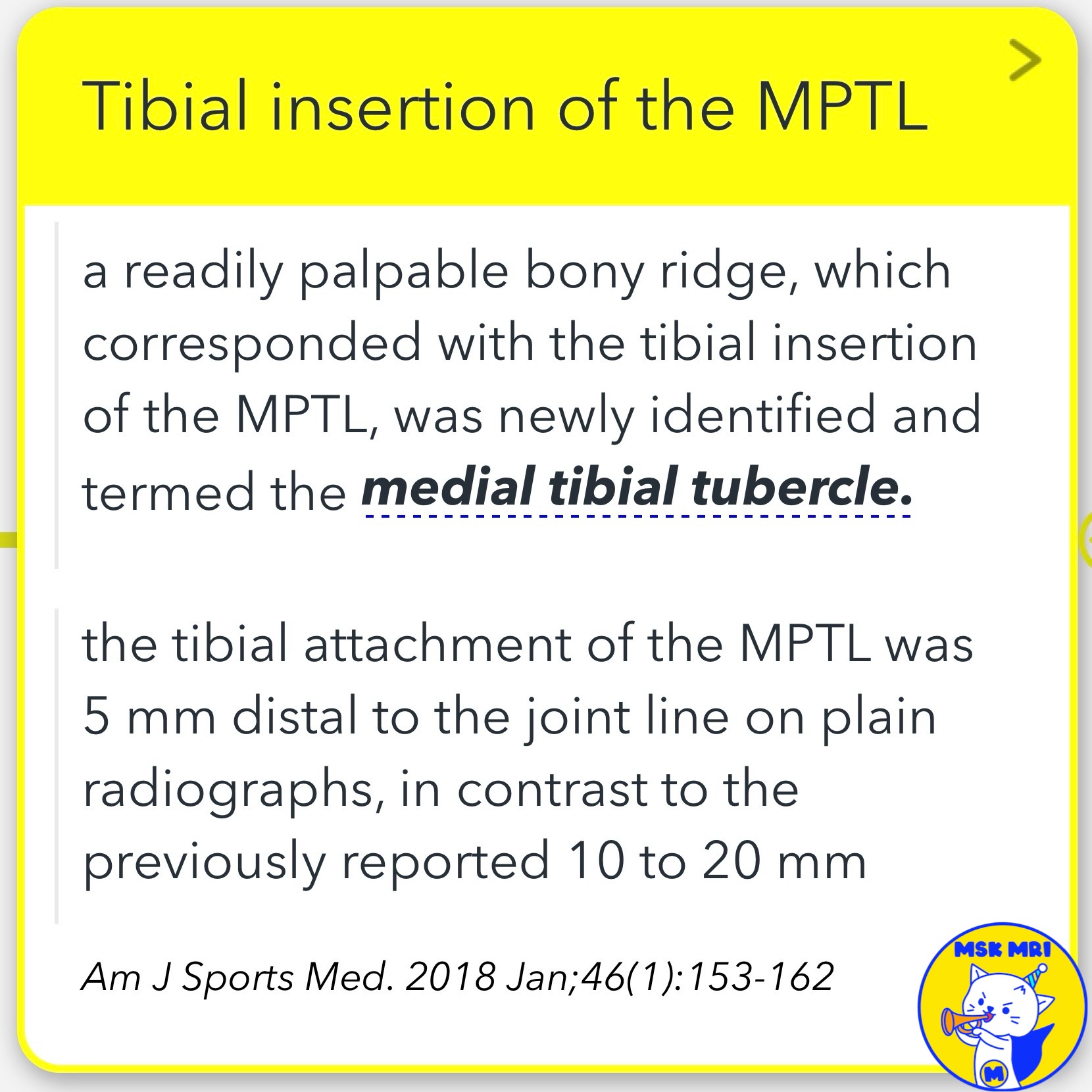
3️⃣ Tibial insertion of the MPTL
- A readily palpable bony ridge, which corresponded with the tibial insertion of the MPTL, was newly identified and termed the medial tibial tubercle.
- The tibial attachment of the MPTL was 5 mm distal to the joint line on plain radiographs, in contrast to the previously reported 10 to 20 mm
📌 Attachments of the medial collateral ligament
1️⃣ sMCL-femoral attachment
- Historically reported to be attached to the medial epicondyle (ME)
- LaPrade et al.: Recent publications suggest the attachment is 3.2mm proximal and 4.8mm posterior to the ME
- Imperial College London: demonstrated that the sMCL covers the ME, with the attachment centered 1-2mm proximal
2️⃣ sMCL-tibial attachment
- A proximal site 10-12mm distal to the joint line (primarily to soft tissues)
- A broader attachment 42-71mm from the joint line
3️⃣ dMCL-femoral attachment
- An important independent stabilizer, despite being adherent to the articular capsule
- Proximal to the sMCL (6mm distal and 5mm posterior to the medial epicondyle)
- 15-17mm above the femoral articular cartilage
4️⃣ dMCL-tibial attachment
- Runs distally from posterior to anterior
- Ends in a fan-wide tibial attachment around 8mm distal to the joint line
- Has two major expansions to the meniscus: meniscofemoral and meniscotibial ligament
Skeletal Radiology (2022) 51:1381–1389
J Bone Joint Surg Am. 2007;89:2000-10
Radiographics. 2023 Jun;43(6):e220177
Arthroscopy. 2017 Oct;33(10):1862-1873
Am J Sports Med. 2018 Jan;46(1):153-162
Knee Surg Sports Traumatol Arthrosc. 2020;28(12)
Orthop Rev (Pavia). 2021 Jun 4;13(2):24463
J Bone Joint Surg Am. 2007;89(9):2000-2010
"Visualizing MSK Radiology: A Practical Guide to Radiology Mastery"
© 2022 MSK MRI Jee Eun Lee All Rights Reserved.
No unauthorized reproduction, redistribution, or use for AI training.
#kneeanatomy, #medialkneestructures, #mpfl, #addductortubercle, #mcl, #patellarligaments, #tibialinsertions, #femoralattachments, #kneestabilizers, #orthoanatomy


https://www.amazon.com/dp/B0DJGMHMFS