Click the link to purchase on Amazon 🎉📚
==============================================
🎥 Check Out All Videos at Once! 📺
👉 Visit Visualizing MSK Blog to explore a wide range of videos! 🩻
https://visualizingmsk.blogspot.com/?view=magazine
📚 You can also find them on MSK MRI Blog and Naver Blog! 📖
https://www.instagram.com/msk_mri/
Click now to stay updated with the latest content! 🔍✨
==============================================
📌 Dejour et al. Classification of Trochlear Dysplasia
1️⃣ Type A
- Signs: Crossing sign laterally
- Axial View: Sulcus angle >145° (shallow trochlea)
- Description: The crossing sign is the only present sign. On axial views, the trochlea is shallower than normal.
2️⃣ Type B
- Signs: Crossing sign and supratrochlear spur laterally
- Axial View: Flat or convex trochlea
- Description: The crossing sign and the supratrochlear spur are present. On axial views, the trochlea is flat.
3️⃣ Type C
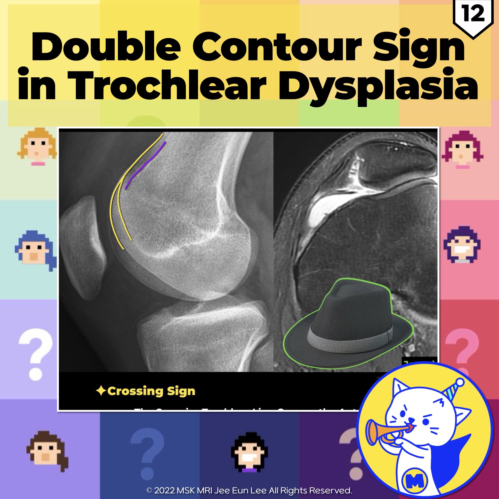
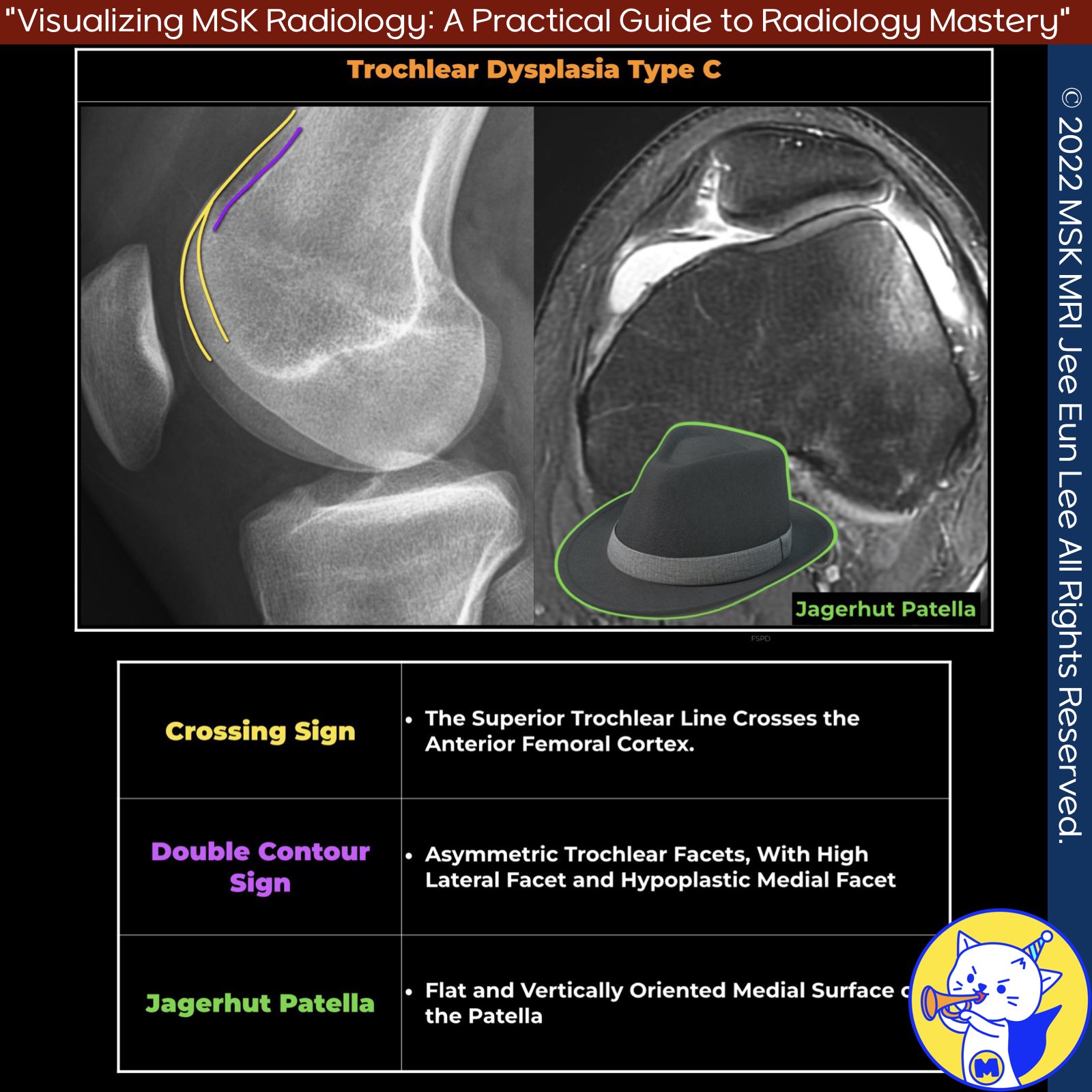
- Signs: Crossing sign and double contour sign laterally
- Axial View: Asymmetry of femoral condyles with hypoplastic medial condyle
- Description: The crossing sign and the double contour sign are present, but there is no spur. On axial views, the medial facet is hypoplastic.
4️⃣ Type D
- Signs: Crossing sign, supratrochlear spur, and double contour sign laterally
- Axial View: Asymmetry of condyles with cliff pattern
- Description: Combines the three signs: crossing sign, supratrochlear spur, and double contour. On axial views, there is a cliff pattern.
➡️ References:
Knee Surg Relat Res. 2023 Mar 13;35(1):7
Clin Sports Med. 2014 Jul;33(3):413-36
MRI Web Clinic – June 2021 Tissue Delamination
"Visualizing MSK Radiology: A Practical Guide to Radiology Mastery"
© 2022 MSK MRI Jee Eun Lee All Rights Reserved.
No unauthorized reproduction, redistribution, or use for AI training.
#trochleardysplasia, #crossingsign, #doublecontoursign, #dejourcriteria, #femoraldeformity, #patelladeformity, #jagerhutdeformity,
'✅ Knee MRI Mastery > Chap 4A. Patelloefemoral joint' 카테고리의 다른 글
| (Fig 4-A.14) Trochlear Dysplasia Assessment Measurements: Part 1 (0) | 2024.06.02 |
|---|---|
| (Fig 4-A.13) Supratrochlear Spur in Trochlear Dysplasia (1) | 2024.06.02 |
| (Fig 4-A.11) Crossing Sign in Trochlear Dysplasia (0) | 2024.06.01 |
| (Fig 4-A.10) Lateral Retinacular Complex (0) | 2024.06.01 |
| (Fig 4-A.09) Medial Patellotibial Ligament (MPTL)/ Sagittal image (0) | 2024.05.31 |


