Click the link to purchase on Amazon 🎉📚
==============================================
🎥 Check Out All Videos at Once! 📺
👉 Visit Visualizing MSK Blog to explore a wide range of videos! 🩻
https://visualizingmsk.blogspot.com/?view=magazine
📚 You can also find them on MSK MRI Blog and Naver Blog! 📖
https://www.instagram.com/msk_mri/
Click now to stay updated with the latest content! 🔍✨
==============================================
📌 Trochlear Dysplasia and the Crossing Sign
✅ Definition of Trochlear Dysplasia
- Trochlear dysplasia refers to an abnormal shape of the trochlear groove, where the patella sits, often appearing shallow or short.
✅ The Crossing Sign
- On lateral radiographs, the presence of trochlear dysplasia can be identified by the "crossing sign," where the trochlear groove line (sulcus line) crosses or lies anterior to the lines formed by the femoral condyles.
✅ Dejour et al. Classification of Trochlear Dysplasia
1️⃣ Type A
- Signs: Crossing sign laterally
- Axial View: Sulcus angle >145° (shallow trochlea)
- Description: The crossing sign is the only present sign. On axial views, the trochlea is shallower than normal.
2️⃣ Type B
- Signs: Crossing sign and supratrochlear spur laterally
- Axial View: Flat or convex trochlea
- Description: The crossing sign and the supratrochlear spur are present. On axial views, the trochlea is flat.
3️⃣ Type C
- Signs: Crossing sign and double contour sign laterally
- Axial View: Asymmetry of femoral condyles with hypoplastic medial condyle
- Description: The crossing sign and the double contour sign are present, but there is no spur. On axial views, the medial facet is hypoplastic.
4️⃣ Type D
- Signs: Crossing sign, supratrochlear spur, and double contour sign laterally
- Axial View: Asymmetry of condyles with cliff pattern
- Description: Combines the three signs: crossing sign, supratrochlear spur, and double contour. On axial views, there is a cliff pattern.
✅ Imaging Assessment
Axial or Merchant view radiographs and MRI provide more accurate evaluation of patellar morphology and trochlear sulcus angle.
- Sulcus angle > 145° with the knee at 30° flexion is consistent with trochlear dysplasia.
- MRI can reliably grade the proximal trochlea as normal, mildly, moderately, or severely dysplastic, guiding management decisions.
➡️ References:
- Skeletal Radiology (2019) 48:859–869
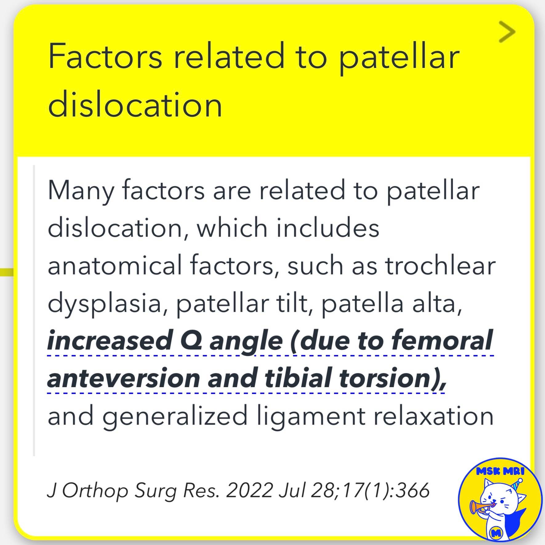
- J Orthop Surg Res. 2022 Jul 28;17(1):366
- Radiographics. 2023 Jun;43(6):e220177
- Knee Surg Relat Res. 2023 Mar 13;35(1):7
"Visualizing MSK Radiology: A Practical Guide to Radiology Mastery"
© 2022 MSK MRI Jee Eun Lee All Rights Reserved.
No unauthorized reproduction, redistribution, or use for AI training.
#TrochlearDysplasia, #CrossingSign, #PatellarInstability, #PatellarDislocation, #DejourClassification, #SulcusAngle, #MRIGrading, #PatellofemoralJoint, #KneeImaging, #OrthopedicRadiology


'✅ Knee MRI Mastery > Chap 4A. Patelloefemoral joint' 카테고리의 다른 글
| (Fig 4-A.13) Supratrochlear Spur in Trochlear Dysplasia (1) | 2024.06.02 |
|---|---|
| (Fig 4-A.12) Double Contour sign in Trochlear Dysplasia (0) | 2024.06.01 |
| (Fig 4-A.10) Lateral Retinacular Complex (0) | 2024.06.01 |
| (Fig 4-A.09) Medial Patellotibial Ligament (MPTL)/ Sagittal image (0) | 2024.05.31 |
| (Fig 4-A.08) Medial Patellar Retinacular Complex: Axial image 4 (MPML and MPTL) (1) | 2024.05.31 |