https://youtu.be/vzxclU_34OI?si=B88NykUJHSELzyAC
Click the link to purchase on Amazon 🎉📚
==============================================
🎥 Check Out All Videos at Once! 📺
👉 Visit Visualizing MSK Blog to explore a wide range of videos! 🩻
https://visualizingmsk.blogspot.com/?view=magazine
📚 You can also find them on MSK MRI Blog and Naver Blog! 📖
https://www.instagram.com/msk_mri/
Click now to stay updated with the latest content! 🔍✨
==============================================
📌 Five patellofemoral joint instability indices for assessing the trochlear dysplasia
1️⃣ Sulcus Angle (SA):
- Measures the angle between the lines defining the lateral and medial trochlear facets.
- A normal SA is around 145°. Higher values indicate pathological flattening.
2️⃣ Trochlear Facet Asymmetry (TFA):
- Ratio between the lengths of the medial and lateral trochlear facets.
- A TFA less than 40% indicates dysplasia.
- Measured 3 cm above the femorotibial joint space.
3️⃣ Lateral Trochlear Inclination (LTI):
- Angle between the subchondral bone of the lateral trochlear facet and the posterior femoral condylar line.
- An LTI angle of less than 11° is considered abnormal.
- Assessed on the first craniocaudal image showing trochlear cartilage.
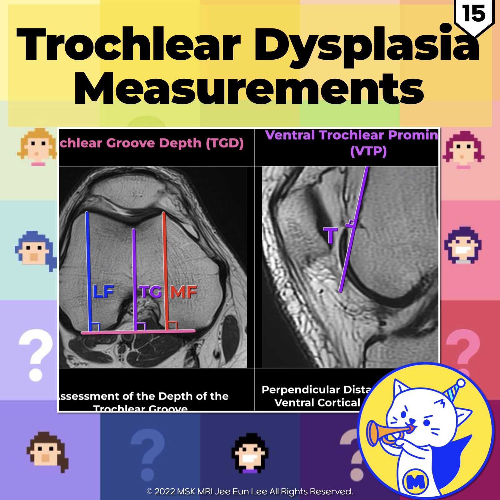
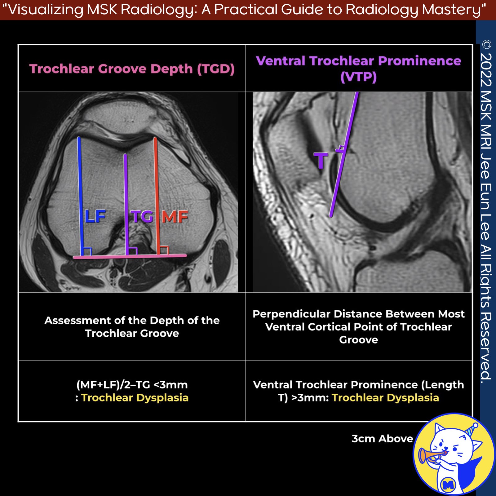
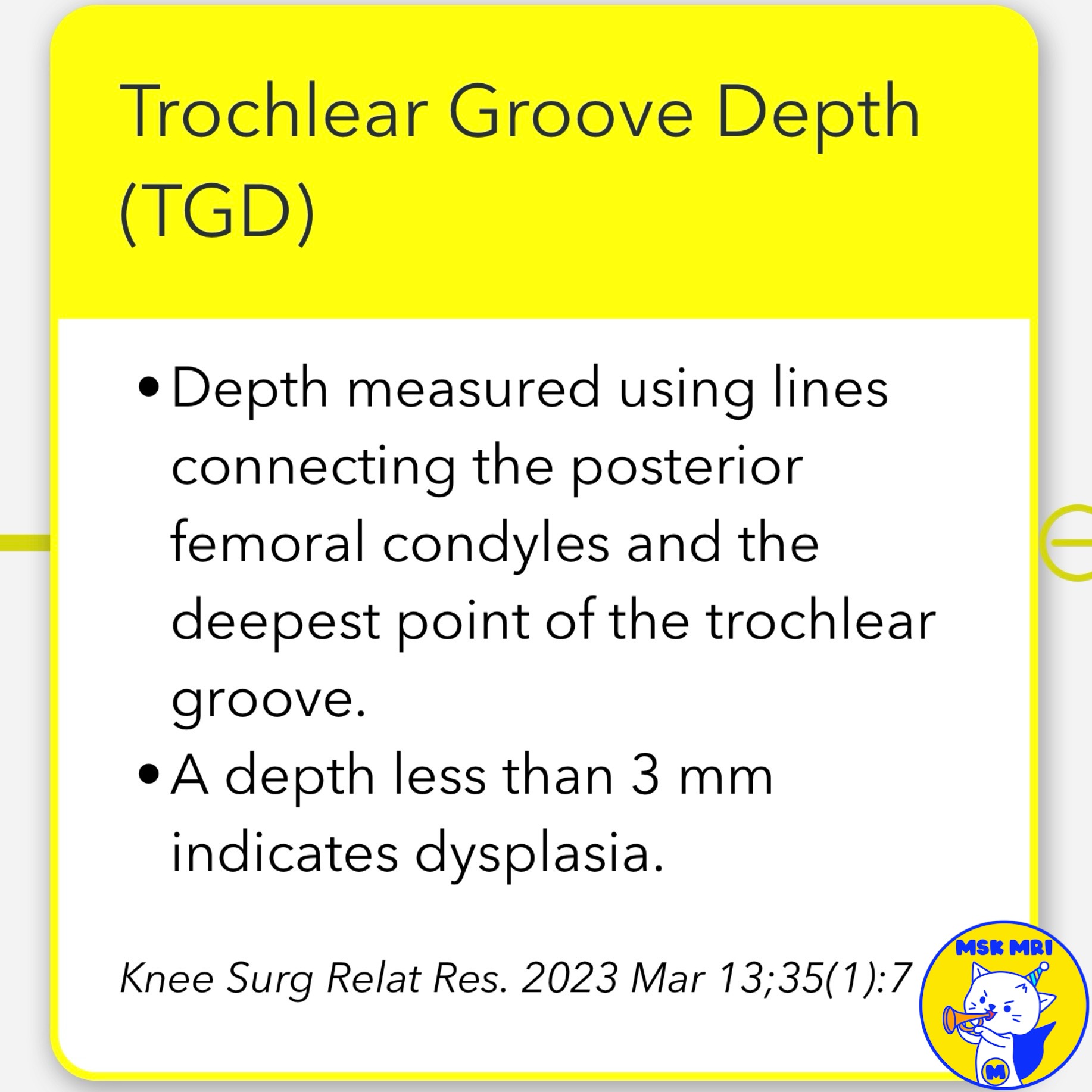
4️⃣ Trochlear Groove Depth (TGD):
- Depth measured using lines connecting the posterior femoral condyles and the deepest point of the trochlear groove.
- A depth less than 3 mm indicates dysplasia.
5️⃣ Ventral Trochlear Prominence (VTP):
- Perpendicular distance between the most ventral point of the trochlear groove and a line parallel to the ventral cortical surface of the distal femur.
- Pathologic threshold values vary; commonly cited values are 3 mm (Dejour et al.) and 8 mm (Pfirrmann et al.).
➡️ References:
Knee Surg Relat Res. 2023 Mar 13;35(1):7
"Visualizing MSK Radiology: A Practical Guide to Radiology Mastery"
© 2022 MSK MRI Jee Eun Lee All Rights Reserved.
No unauthorized reproduction, redistribution, or use for AI training.
#TrochlearDysplasia, #PatellofemoralInstability, #SulcusAngle, #TrochlearFacetAsymmetry, #LateralTrochlearInclination, #TrochlearGrooveDepth, #VentralTrochlearProminence, #OrthopedicRadiology, #KneeMRI,


'✅ Knee MRI Mastery > Chap 4A. Patelloefemoral joint' 카테고리의 다른 글
| (Fig 4-A.17) Patellar Height Measurements: Part 1 (0) | 2024.06.02 |
|---|---|
| (Fig 4-A.16) Patella Alta and Patella Baja (2) | 2024.06.02 |
| (Fig 4-A.14) Trochlear Dysplasia Assessment Measurements: Part 1 (0) | 2024.06.02 |
| (Fig 4-A.13) Supratrochlear Spur in Trochlear Dysplasia (1) | 2024.06.02 |
| (Fig 4-A.12) Double Contour sign in Trochlear Dysplasia (0) | 2024.06.01 |