https://youtu.be/tOk0dMOGt18?si=lbapoRVI2jXagNPk
https://youtu.be/vzxclU_34OI?si=B88NykUJHSELzyAC
Click the link to purchase on Amazon 🎉📚
==============================================
🎥 Check Out All Videos at Once! 📺
👉 Visit Visualizing MSK Blog to explore a wide range of videos! 🩻
https://visualizingmsk.blogspot.com/?view=magazine
📚 You can also find them on MSK MRI Blog and Naver Blog! 📖
https://www.instagram.com/msk_mri/
Click now to stay updated with the latest content! 🔍✨
==============================================
📌 MPFL Injury in Lateral Patellar Dislocation
✅ Prevalence and Location
- The MPFL is injured in almost all (∼95%) patellar dislocations
- The location of the injury varies: 37% at the patellar attachment, 37% at the femoral attachment, 16% midsubstance, and the remainder at a combination of sites
✅ MRI Findings
- Thickening, intraligamentous fluid, waviness of the fibers, and frank discontinuity indicate MPFL injury
✅ Case Study: Evaluating MPFL Components
1️⃣ Medial Quadriceps Tendon Femoral Ligament Component
- Identified within the purple dotted lines on the proximal axial image
- This patient shows a partial tear compared to normal
2️⃣ Transverse Component
- Highlighted in yellow on the axial image
- Femoral and patellar side partial tear of the transverse component observed
3️⃣ Oblique Decussation Component
- Highlighted in pink, attaches to the proximal superficial MCL fibers
- Femoral and mid-substance partial tear of the oblique decussation component observed
📌 Lateral Patellar Dislocation
✅ MPFL Injury:
- MPFL is injured in ~95% of patellar dislocations
- Injury location varies: 37% patellar attachment, 37% femoral attachment, 16% midsubstance
- MRI findings: thickening, fluid, waviness, discontinuity
✅ Injury Pattern:
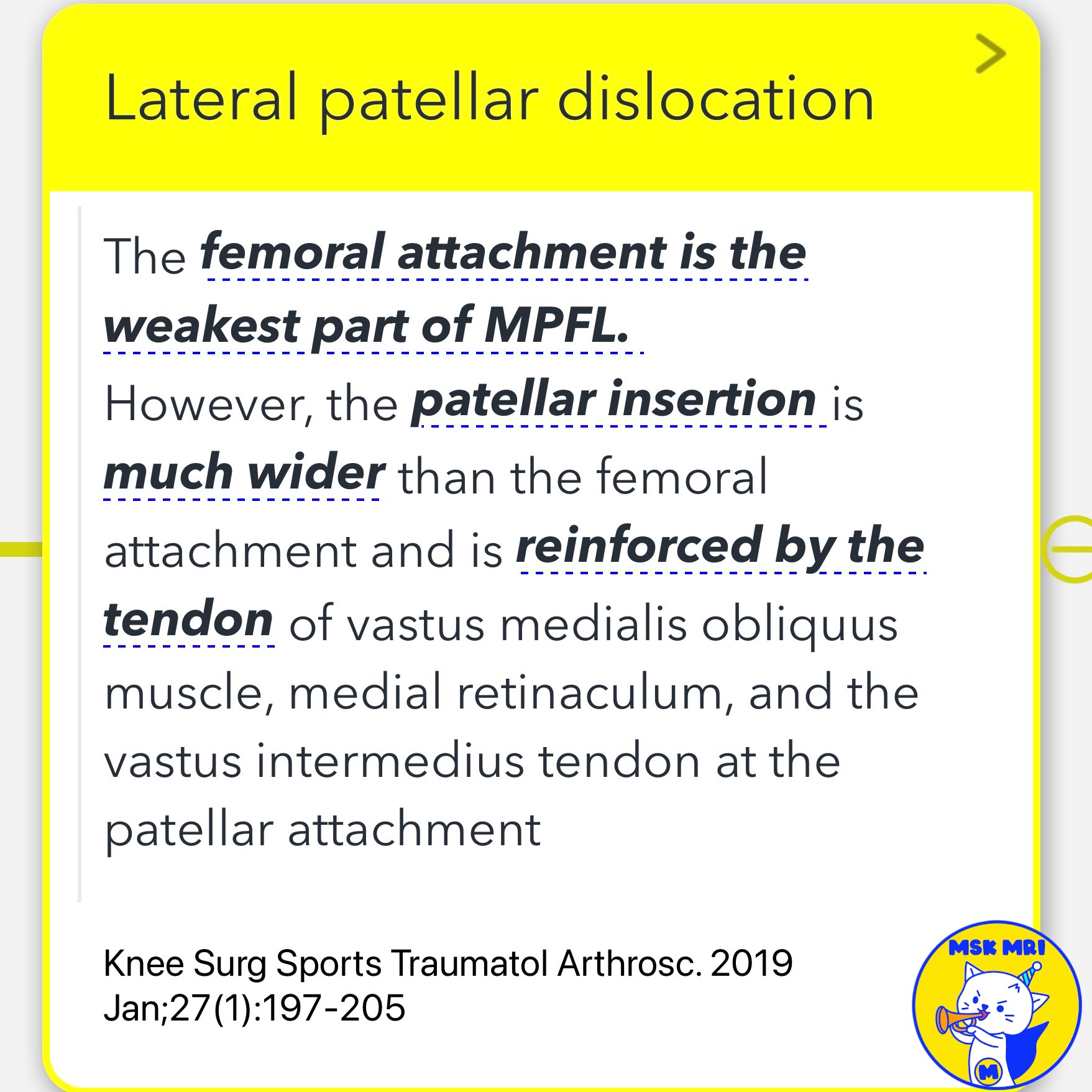
- Femoral attachment is weakest part of MPFL
- Patellar insertion reinforced by surrounding structures
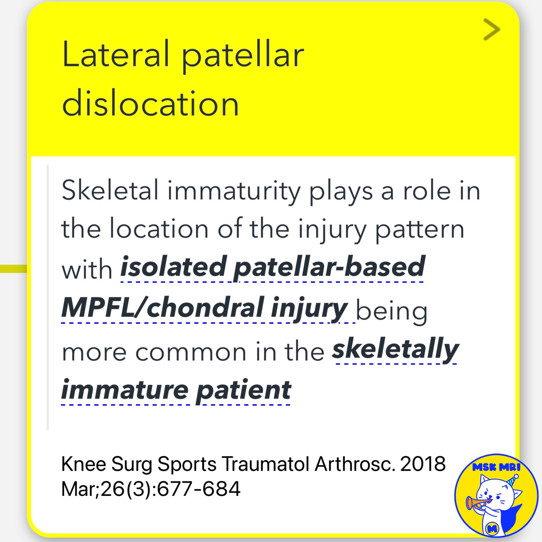
- In skeletally immature, patellar-based MPFL/chondral injury more common
➡️ References:
Radiographics. 2023 Jun;43(6):e220177
RadioGraphics 2018; 38:2069–2101
Knee Surg Sports Traumatol Arthrosc. 2019 Jan;27(1):197-205
Knee Surg Sports Traumatol Arthrosc. 2018 Mar;26(3):677-684
"Visualizing MSK Radiology: A Practical Guide to Radiology Mastery"
© 2022 MSK MRI Jee Eun Lee All Rights Reserved.
No unauthorized reproduction, redistribution, or use for AI training.
#LateralPatellarDislocation, #MPFL, #MRIFindings, #QuadricepsTendonFemoralLigament, #TransverseComponent, #ObliqueDecussationComponent, #PartialTear, #MCL, #PatelloFemoralLigament, #KneeInjury


'✅ Knee MRI Mastery > Chap 4A. Patelloefemoral joint' 카테고리의 다른 글
| (Fig 4-A.31) Three Types of Patellar MPFL Injuries at the Patella (2) | 2024.06.05 |
|---|---|
| (Fig 4-A.30) Complete MPFL Tear at Femoral Origin (0) | 2024.06.05 |
| (Fig 4-A.28) Displaced Osteochondral Fragments (0) | 2024.06.04 |
| (Fig 4-A.27) Headless Compression Screw Fixation of Osteochondral Fracture (0) | 2024.06.04 |
| (Fig 4-A.26) Displaced Osteochondral Fragments in Patellar Dislocation (0) | 2024.06.04 |