https://youtu.be/vzxclU_34OI?si=B88NykUJHSELzyAC
==============================================
⬇️✨⬇️🎉⬇️🔥⬇️📚⬇️
Click the link to purchase on Amazon 🎉📚
==============================================
🎥 Check Out All Videos at Once! 📺
👉 Visit Visualizing MSK Blog to explore a wide range of videos! 🩻
https://visualizingmsk.blogspot.com/?view=magazine
📚 You can also find them on MSK MRI Blog and Naver Blog! 📖
https://www.instagram.com/msk_mri/
Click now to stay updated with the latest content! 🔍✨
==============================================
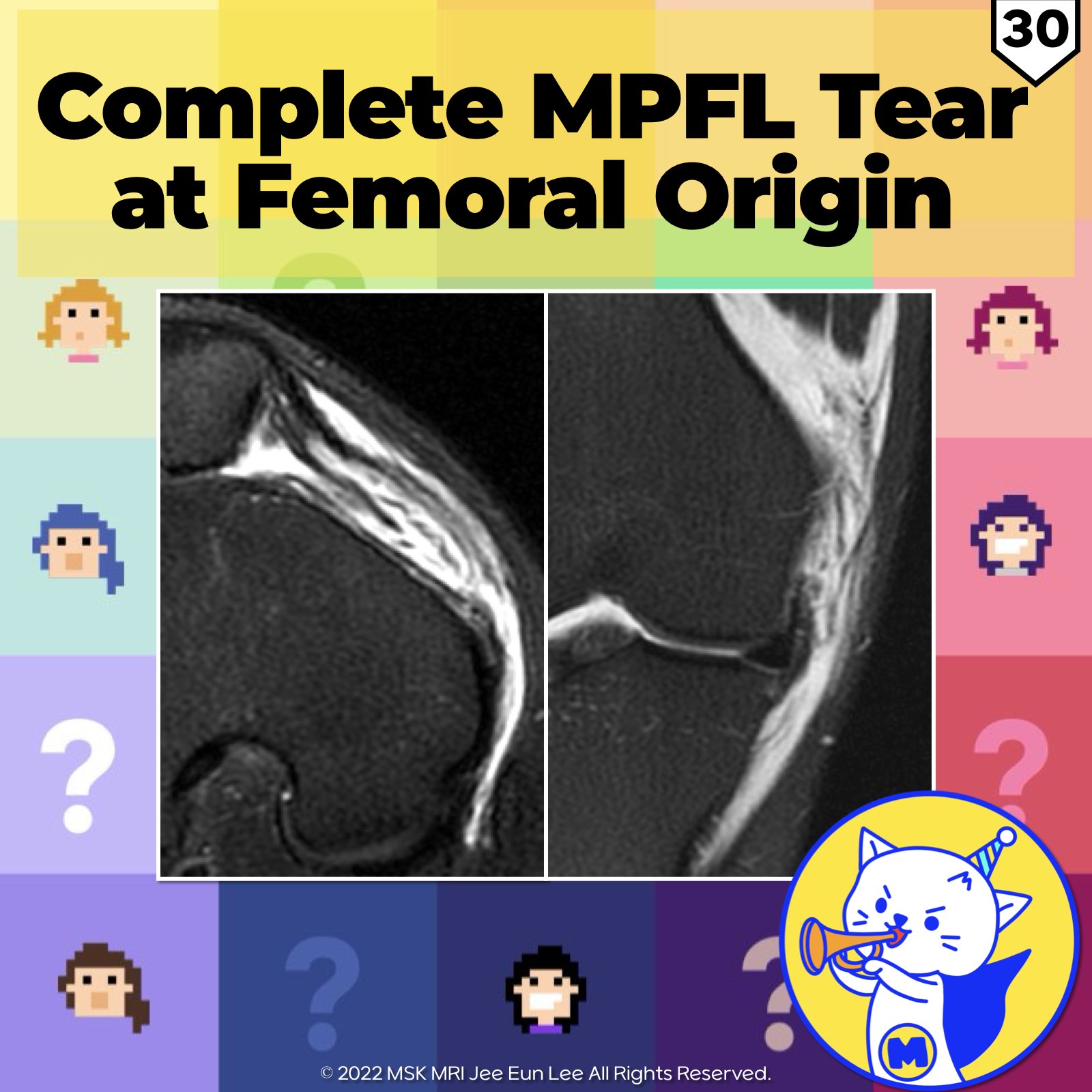
📌 Complete MPFL Tear at Femoral Origin
- Lateral patellar dislocation often involves the medial patellofemoral ligament (MPFL), which has its weakest attachment at the femoral side.
- The patellar insertion of the MPFL is much wider and reinforced by the vastus medialis obliquus muscle, medial retinaculum, and the vastus intermedius tendon.
- The femoral origin is the most common site of injury, which often results in a decreased likelihood of returning to pre-injury activity levels.
References:
- Knee Surg Sports Traumatol Arthrosc. 2019 Jan;27(1):197-205.
- Skeletal Radiol (2012) 41:137–148.
"Visualizing MSK Radiology: A Practical Guide to Radiology Mastery"
© 2022 MSK MRI Jee Eun Lee All Rights Reserved.
No unauthorized reproduction, redistribution, or use for AI training.
#MPFL #patellardislocation #kneesurgery #sportsinjury #orthopedics #radiology #kneetreatment #femoralattachment #patellainjury #medialretinaculum


'✅ Knee MRI Mastery > Chap 4A. Patelloefemoral joint' 카테고리의 다른 글
| (Fig 4-A.32) Type P0/ Ligament Disruption at Patellar Attachment (0) | 2024.06.05 |
|---|---|
| (Fig 4-A.31) Three Types of Patellar MPFL Injuries at the Patella (2) | 2024.06.05 |
| (Fig 4-A.29) Partial MPFL Tear at Multiple Sites (0) | 2024.06.05 |
| (Fig 4-A.28) Displaced Osteochondral Fragments (0) | 2024.06.04 |
| (Fig 4-A.27) Headless Compression Screw Fixation of Osteochondral Fracture (0) | 2024.06.04 |