https://youtu.be/vzxclU_34OI?si=B88NykUJHSELzyAC
Click the link to purchase on Amazon 🎉📚
==============================================
🎥 Check Out All Videos at Once! 📺
👉 Visit Visualizing MSK Blog to explore a wide range of videos! 🩻
https://visualizingmsk.blogspot.com/?view=magazine
📚 You can also find them on MSK MRI Blog and Naver Blog! 📖
https://www.instagram.com/msk_mri/
Click now to stay updated with the latest content! 🔍✨
==============================================
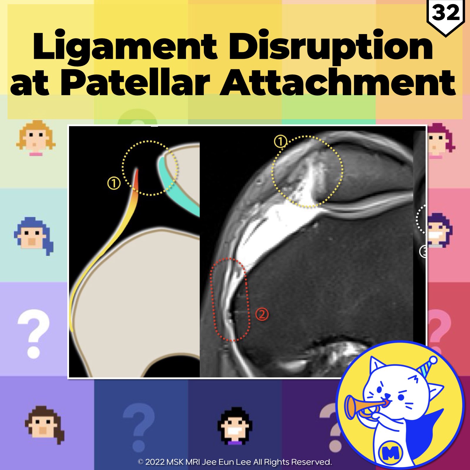
📌 Ligament Disruption at Patellar Attachment
- The patellar insertion is wider and reinforced by surrounding tendons and retinacula.
- Ruptures at this site or the MPFL midsubstance without avulsion fragment are generally not associated with significant patellar instability. (Reference: Knee Surg Sports Traumatol Arthrosc (2014) 22:2414–2418)
✅Femoral MPFL Attachment
- The femoral attachment is the weakest part of the MPFL.
- The patellar insertion is much wider and reinforced by the tendon of the vastus medialis obliquus muscle, medial retinaculum, and the vastus intermedius tendon. (Reference: Knee Surg Sports Traumatol Arthrosc. 2019 Jan;27(1):197-205)
✅ Transverse Component of the MPFL
- The femoral attachment of the tMPFL is assessed immediately below the adductor tubercle and above the superior attachment of the sMCL.
- It is very thin and difficult to visualize even in the uninjured state.
- Readers characterized this femoral attachment as "no edema" or "edema" rather than using a grading system. (Reference: Skeletal Radiology (2022) 51:1381–1389)
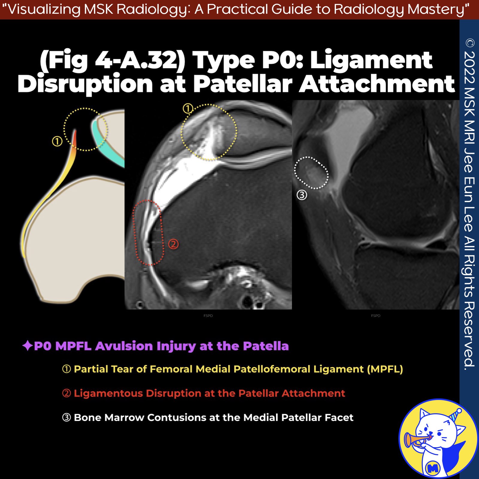
✅Case MRI Findings
- Ligamentous partial disruption at the patellar attachment
- Bone marrow contusions at the medial patellar facet
- No bony avulsion fracture from the medial patella
- Edema noted at the femoral attachment of the MPFL, indicating a partial tear of the femoral MPFL component.
"Visualizing MSK Radiology: A Practical Guide to Radiology Mastery"
© 2022 MSK MRI Jee Eun Lee All Rights Reserved.
No unauthorized reproduction, redistribution, or use for AI training.
#MPFLinjury, #PatellarAvulsion, #FemoralAttachment, #BoneMarrowContusion, #LigamentDisruption, #PatellarInstability, #MPFLTear, #MRIFindings, #OrthopaedicSurgery, #SportsMedicine


'✅ Knee MRI Mastery > Chap 4A. Patelloefemoral joint' 카테고리의 다른 글
| (Fig 4-A.34) Type P2/ Bony Avulsion with Articular Cartilage from Medial Patella (0) | 2024.06.06 |
|---|---|
| (Fig 4-A.33) Type P1: Bony Avulsion Fracture from Medial Patella (0) | 2024.06.05 |
| (Fig 4-A.31) Three Types of Patellar MPFL Injuries at the Patella (2) | 2024.06.05 |
| (Fig 4-A.30) Complete MPFL Tear at Femoral Origin (0) | 2024.06.05 |
| (Fig 4-A.29) Partial MPFL Tear at Multiple Sites (0) | 2024.06.05 |