Click the link to purchase on Amazon 🎉📚
==============================================
🎥 Check Out All Videos at Once! 📺
👉 Visit Visualizing MSK Blog to explore a wide range of videos! 🩻
https://visualizingmsk.blogspot.com/?view=magazine
📚 You can also find them on MSK MRI Blog and Naver Blog! 📖
https://www.instagram.com/msk_mri/
Click now to stay updated with the latest content! 🔍✨
==============================================
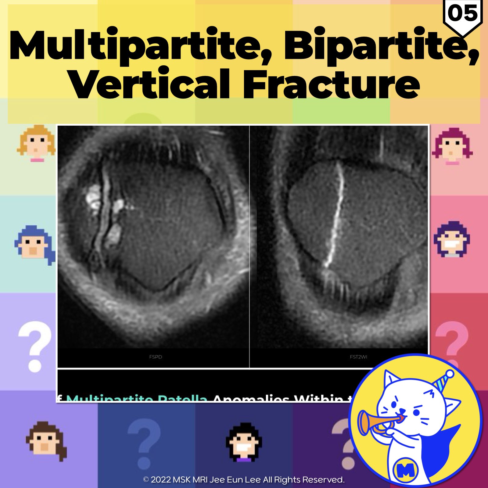
📌 Differentiating Bipartite and Multipartite Patella from Fractures
- A bipartite or multipartite patella, which involves the presence of more than one ossicle or bone fragment, may be mistaken for a fracture. However, it can usually be distinguished from a fracture by the following features:
1️⃣ Typical Location:
- Bipartite and multipartite patellae tend to occur in specific locations, such as the superolateral pole or lateral margin.
2️⃣ Well-Corticated Margins:
The ossicles or fragments in bipartite and multipartite patellae often have well-corticated margins, indicating a developmental process rather than a recent fracture.
3️⃣ Articular Cartilage Integrity:
The articular cartilage of the bipartite segment remains intact, unlike in fractures where the cartilage may be disrupted.
4️⃣ Normal Marrow Signal:
- The ossicles or fragments should exhibit normal marrow signal, unlike fracture fragments, which may show bone marrow edema or abnormal signal.
5️⃣ Smooth Synchondrosis:
- The synchondrosis, or the junction between the ossicles, should appear smooth and show normal cartilage signal.
6️⃣ Osseous or Fibrous Union:
- Bipartite and multipartite patellae may demonstrate osseous or fibrous union between the ossicles, which is not seen in fractures.
7️⃣ Hypertrophy of Unfused Fragments:
- Accessory ossification centers in multipartite patellae can be distinguished from fracture fragments by recognizing the hypertrophy of the unfused fragments and their corticated margins.
8️⃣ Fragment Size:
- In bipartite patellae, the bipartite segment is often larger than expected for a fracture fragment.
References:
- Skeletal Radiol. 2018 Aug;47(8):1069-1086
- Magn Reson Imaging Clin N Am 22 (2014) 601–620
- RadioGraphics 2018; 38:2069–2101
"Visualizing MSK Radiology: A Practical Guide to Radiology Mastery"
© 2022 MSK MRI Jee Eun Lee All Rights Reserved.
No unauthorized reproduction, redistribution, or use for AI training.
#BipartitePatella, #MultipartitePatella, #PatellarFracture, #OssicleHypertrophy, #CorticatedMargins, #CartilageIntegrity, #NormalMarrowSignal, #SmoothSynchondrosis, #OssificationCenters, #FragmentSize.


'✅ Knee MRI Mastery > Chap 4BCD. Anterior knee' 카테고리의 다른 글
| (Fig 4-B.07) Dorsal Defect of the Patella (0) | 2024.06.11 |
|---|---|
| (Fig 4-B.06) Symptomatic Bipartite Patella (1) | 2024.06.10 |
| (Fig 4-B.04) Bipartite Patella and Saupe Classification (1) | 2024.06.10 |
| (Fig 4-B.03) Retropatellar Articular Surface and Wiberg Classification (0) | 2024.06.10 |
| (Fig 4-B.02) Anatomy of the Multilayered Quadriceps Tendon/ Part 2 (0) | 2024.06.09 |