https://youtu.be/eyuYuvl3wOg?si=Oap54zIN_WM_w-r4
==============================================
⬇️✨⬇️🎉⬇️🔥⬇️📚⬇️
Click the link to purchase on Amazon 🎉📚
==============================================
🎥 Check Out All Videos at Once! 📺
👉 Visit Visualizing MSK Blog to explore a wide range of videos! 🩻
https://visualizingmsk.blogspot.com/?view=magazine
📚 You can also find them on MSK MRI Blog and Naver Blog! 📖
https://www.instagram.com/msk_mri/
Click now to stay updated with the latest content! 🔍✨
==============================================
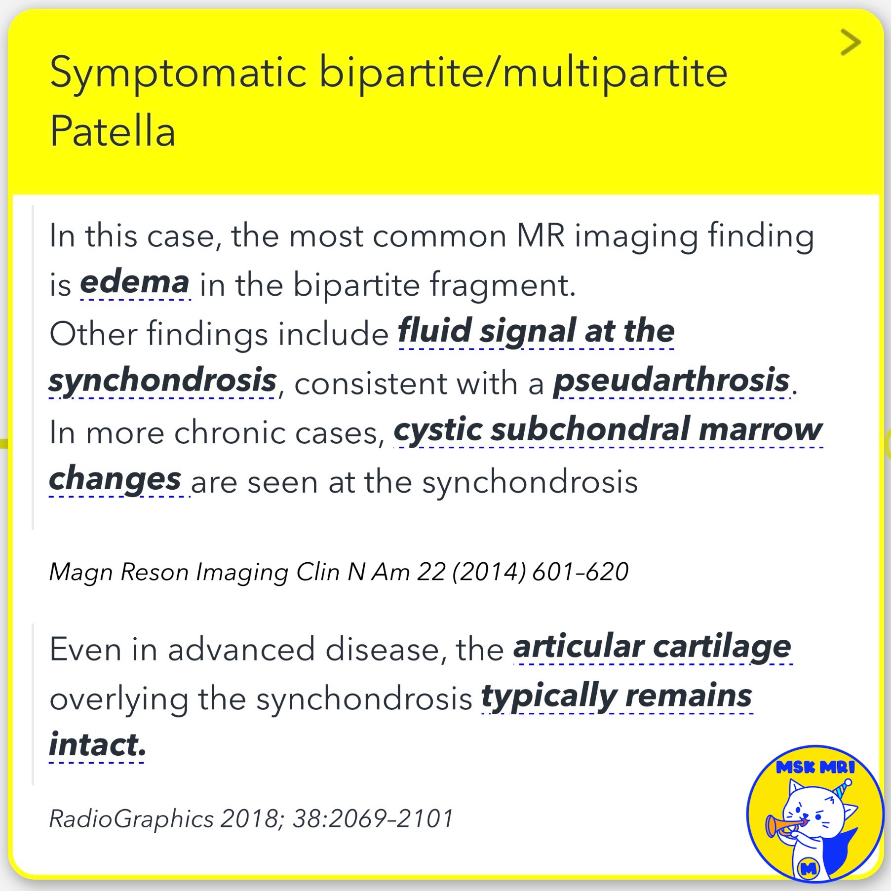
📌 Symptomatic Bipartite or Multipartite Patella
✅ MRI Findings:
- Edema in the bipartite fragment
- Fluid signal at the synchondrosis, suggesting pseudarthrosis
- Cystic subchondral marrow changes at the synchondrosis in chronic cases
- Fluid signal may be seen between the fragment and the patella
✅ Cartilage Involvement:
- Articular cartilage overlying the synchondrosis typically remains intact, even in advanced disease
✅ Treatment:
- Surgical excision or lateral retinacular release for symptomatic bipartite patella
References:
- Skeletal Radiol. 2018 Aug;47(8):1069-1086
- Magn Reson Imaging Clin N Am 22 (2014) 601–620
- RadioGraphics 2018; 38:2069–2101
- Stoller's Orthopaedics and Sports Medicine: by Stoller MD FACR, David W. LWW, 2016
#bipartitepatella, #multipartitepatella, #anteriorkneepainsyndrome, #patellarsyndrome, #MRIfindings, #pseudarthrosis, #subchondralcyst, #articularcartilage, #surgicalexcision, #lateralretinacularrelease.


'✅ Knee MRI Mastery > Chap 4BCD. Anterior knee' 카테고리의 다른 글
| (Fig 4-B.08) Anatomy of Prepatellar Quadriceps Continuation (0) | 2024.06.11 |
|---|---|
| (Fig 4-B.07) Dorsal Defect of the Patella (0) | 2024.06.11 |
| (Fig 4-B.05) Patellar Comparison/ Multipartite, Bipartite, Vertical Fracture (0) | 2024.06.10 |
| (Fig 4-B.04) Bipartite Patella and Saupe Classification (1) | 2024.06.10 |
| (Fig 4-B.03) Retropatellar Articular Surface and Wiberg Classification (0) | 2024.06.10 |