Click the link to purchase on Amazon 🎉📚
==============================================
🎥 Check Out All Videos at Once! 📺
👉 Visit Visualizing MSK Blog to explore a wide range of videos! 🩻
https://visualizingmsk.blogspot.com/?view=magazine
📚 You can also find them on MSK MRI Blog and Naver Blog! 📖
https://www.instagram.com/msk_mri/
Click now to stay updated with the latest content! 🔍✨
==============================================
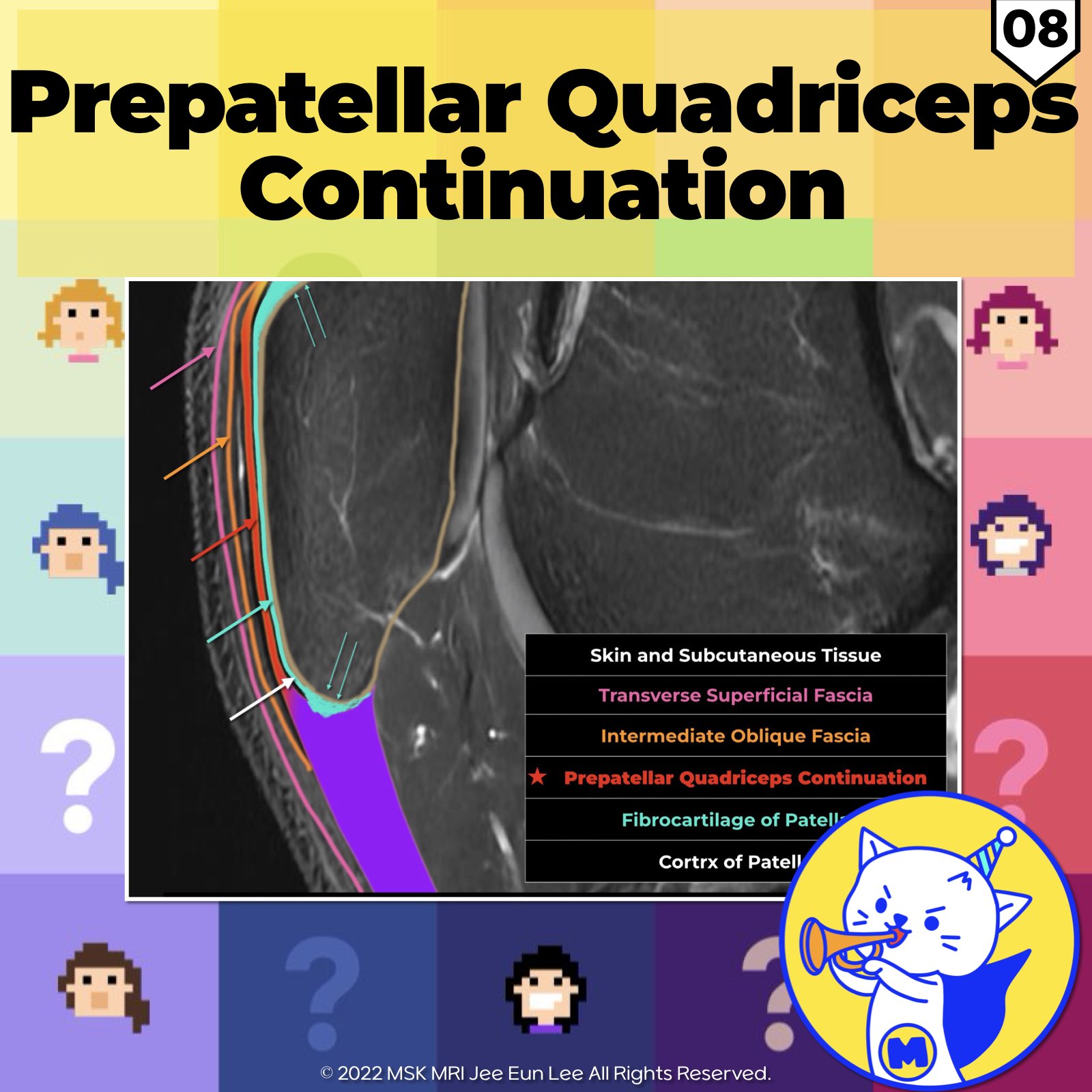
📌The Prepatellar Quadriceps Continuation
- Formed by anterior fibers of rectus femoris tendon
- Inserts onto superior patella, then continues distally
- Adheres to anterior patellar surface via fibrocartilaginous enthesis (#fibrocartilage, #enthesis)
- Joins patellar tendon, inserting onto tibial tuberosity
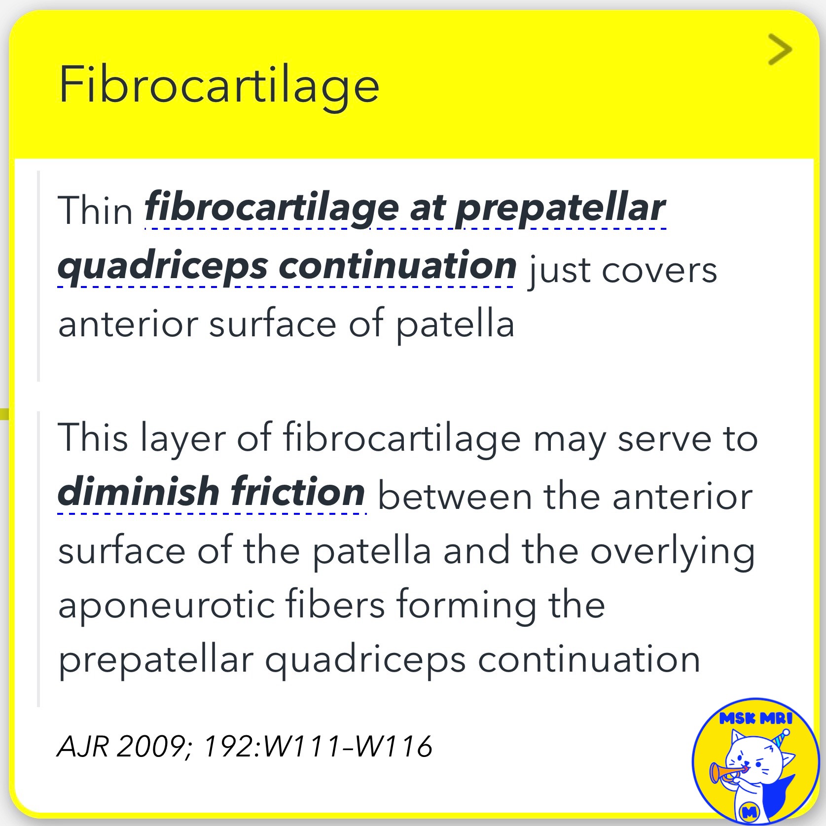
✅ Fibrocartilaginous Attachment
- Thin fibrocartilage layer covers patellar surface at continuation
- Reduces friction against overlying tendon fibers
- Small, loosely attached amount at patellar attachment
- Tendon transitions through zones: dense connective tissue, uncalcified fibrocartilage, calcified fibrocartilage, bone
✅ Imaging and Injury
- Normally no space between continuation and patella
- With injury, thin fluid line may be seen (distinct from bursitis)
- Understanding anatomy aids in evaluating extensor mechanism injuries
References:
RadioGraphics 2018; 38:2069-2101
AJR 2009; 192:W111-W116
Magn Reson Imaging Clin N Am 22 (2014) 601-620
"Visualizing MSK Radiology: A Practical Guide to Radiology Mastery"
© 2022 MSK MRI Jee Eun Lee All Rights Reserved.
No unauthorized reproduction, redistribution, or use for AI training.
#prepatellarquadricepscontinuation, #quadricepstendon, #patellartendon, #fibrocartilage, #enthesis, #tendonboneinterface, #extensorinjury, #patellarinjury, #kneeanatomy, #radiology


'✅ Knee MRI Mastery > Chap 4BCD. Anterior knee' 카테고리의 다른 글
| (Fig 4-B.10) Prepatellar Bursa Compartmentalization (0) | 2024.06.11 |
|---|---|
| (Fig 4-B.09) Prepatellar Soft Tissue Anatomy (2) | 2024.06.11 |
| (Fig 4-B.07) Dorsal Defect of the Patella (0) | 2024.06.11 |
| (Fig 4-B.06) Symptomatic Bipartite Patella (1) | 2024.06.10 |
| (Fig 4-B.05) Patellar Comparison/ Multipartite, Bipartite, Vertical Fracture (0) | 2024.06.10 |