Click the link to purchase on Amazon 🎉📚
==============================================
🎥 Check Out All Videos at Once! 📺
👉 Visit Visualizing MSK Blog to explore a wide range of videos! 🩻
https://visualizingmsk.blogspot.com/?view=magazine
📚 You can also find them on MSK MRI Blog and Naver Blog! 📖
https://www.instagram.com/msk_mri/
Click now to stay updated with the latest content! 🔍✨
==============================================
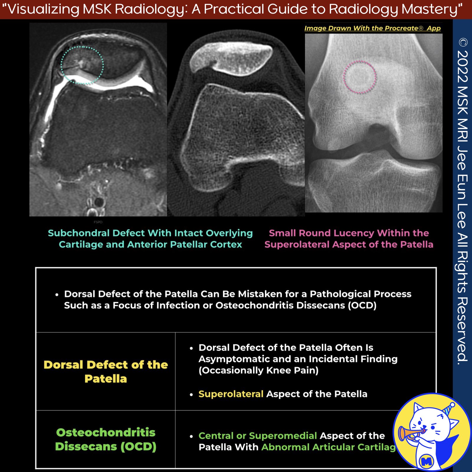
📌Dorsal Defect of the Patella
- The dorsal defect of the patella is generally considered a developmental anomaly of the epiphysis with delayed ossification.
- Despite its unclear (probably complex and multifactorial) etiology, the dorsal patellar defect does not grow in size and usually heals spontaneously with sclerosis, thus requiring no further intervention.
✅ Radiographic Appearance
- Lucent lesion with well-defined sclerotic borders at the superolateral aspect of the patella's dorsal articular surface
✅ MRI Appearance
- Well-defined lesion at the superolateral aspect of the dorsal patella
- Sclerotic margins with heterogeneous internal signal (isointense to hyperintense to cartilage)
- Intact overlying cartilage
✅ Symptomatic Lesions
Symptomatic dorsal defects may exhibit chondral irregularity and marrow edema.
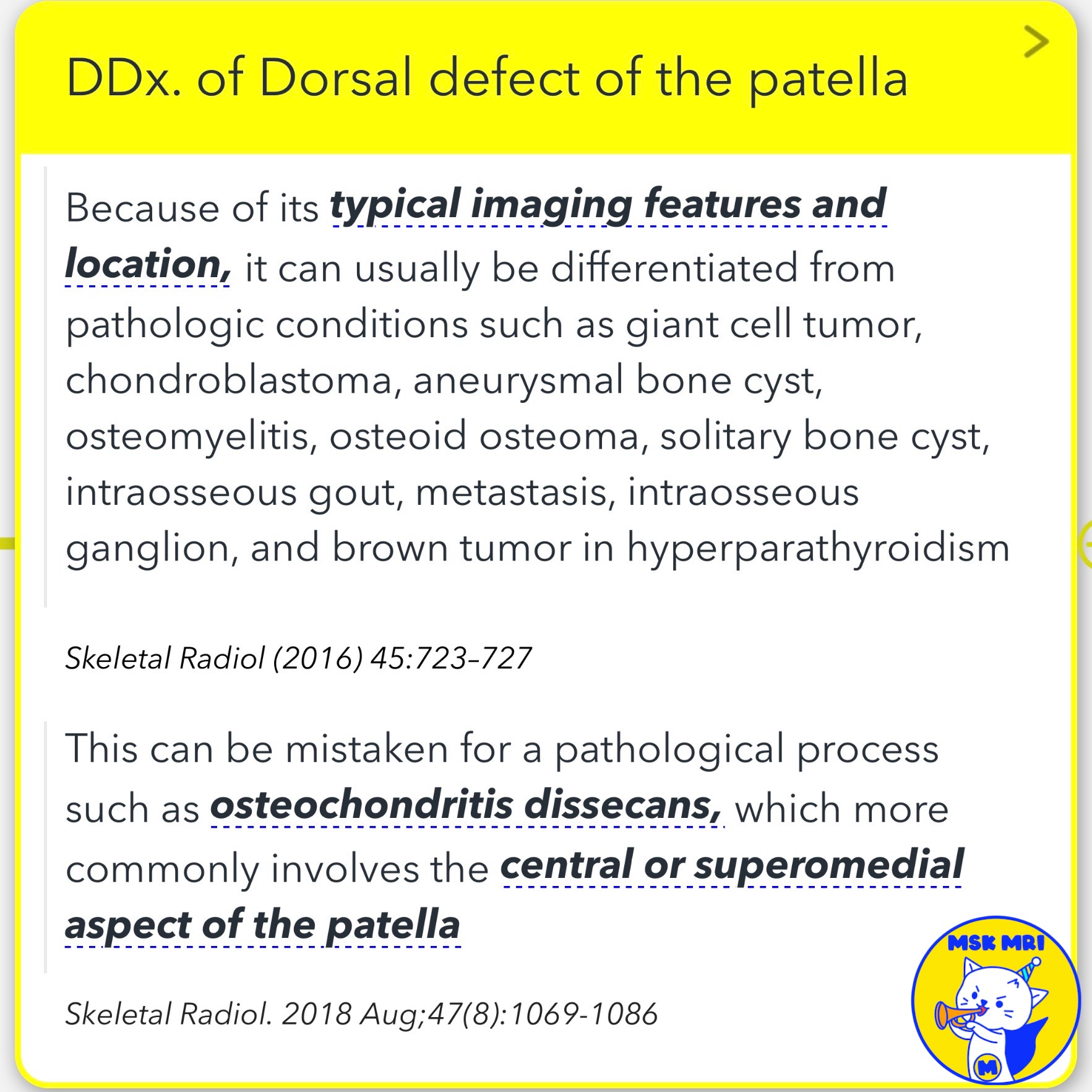
✅ Differential Diagnosis
- Osteochondral injury
- Brodie's abscess
- Osteochondritis dissecans
- Neoplastic bony lesions
- Should not be mistaken for conditions like giant cell tumor, chondroblastoma, aneurysmal bone cyst, osteomyelitis, etc.
✅ Differentiating from Osteochondritis Dissecans
- Osteochondritis dissecans has a predilection for the medial facet of the patella
- May have a separated articular cartilage flap in osteochondritis dissecans
- Osteochondritis dissecans more commonly involves the central or superomedial aspect of the patella
References:
Skeletal Radiol (2016) 45:723-727
Magn Reson Imaging Clin N Am 22 (2014) 601-620
RadioGraphics 2018; 38:2069-2101
Stoller's Orthopaedics and Sports Medicine, LWW, 2016
Skeletal Radiol. 2018 Aug;47(8):1069-1086
"Visualizing MSK Radiology: A Practical Guide to Radiology Mastery"
© 2022 MSK MRI Jee Eun Lee All Rights Reserved.
No unauthorized reproduction, redistribution, or use for AI training.
#DorsalDefectPatella, #DevelopmentalAnomaly, #DelayedOssification, #SclerolicLesion, #ChondralIrregularity, #MarrowEdema, #OsteochondritisDissecans, #DifferentialDiagnosis, #MusculoskeletalRadiology, #OrthopedicsRadiology


'✅ Knee MRI Mastery > Chap 4BCD. Anterior knee' 카테고리의 다른 글
| (Fig 4-B.09) Prepatellar Soft Tissue Anatomy (2) | 2024.06.11 |
|---|---|
| (Fig 4-B.08) Anatomy of Prepatellar Quadriceps Continuation (0) | 2024.06.11 |
| (Fig 4-B.06) Symptomatic Bipartite Patella (1) | 2024.06.10 |
| (Fig 4-B.05) Patellar Comparison/ Multipartite, Bipartite, Vertical Fracture (0) | 2024.06.10 |
| (Fig 4-B.04) Bipartite Patella and Saupe Classification (1) | 2024.06.10 |