Click the link to purchase on Amazon 🎉📚
==============================================
🎥 Check Out All Videos at Once! 📺
👉 Visit Visualizing MSK Blog to explore a wide range of videos! 🩻
https://visualizingmsk.blogspot.com/?view=magazine
📚 You can also find them on MSK MRI Blog and Naver Blog! 📖
https://www.instagram.com/msk_mri/
Click now to stay updated with the latest content! 🔍✨
==============================================
📌Anatomy of the Prepatellar Bursae
✅ Trilaminar Arrangement
- Superficial transverse fascial layer
- Intermediate oblique aponeurotic layer
- Longitudinally oriented fibers of the rectus femoris tendon
✅ Layers and Bursae
- Skin
- Prepatellar subcutaneous bursa
- Superficial transverse fascial layer
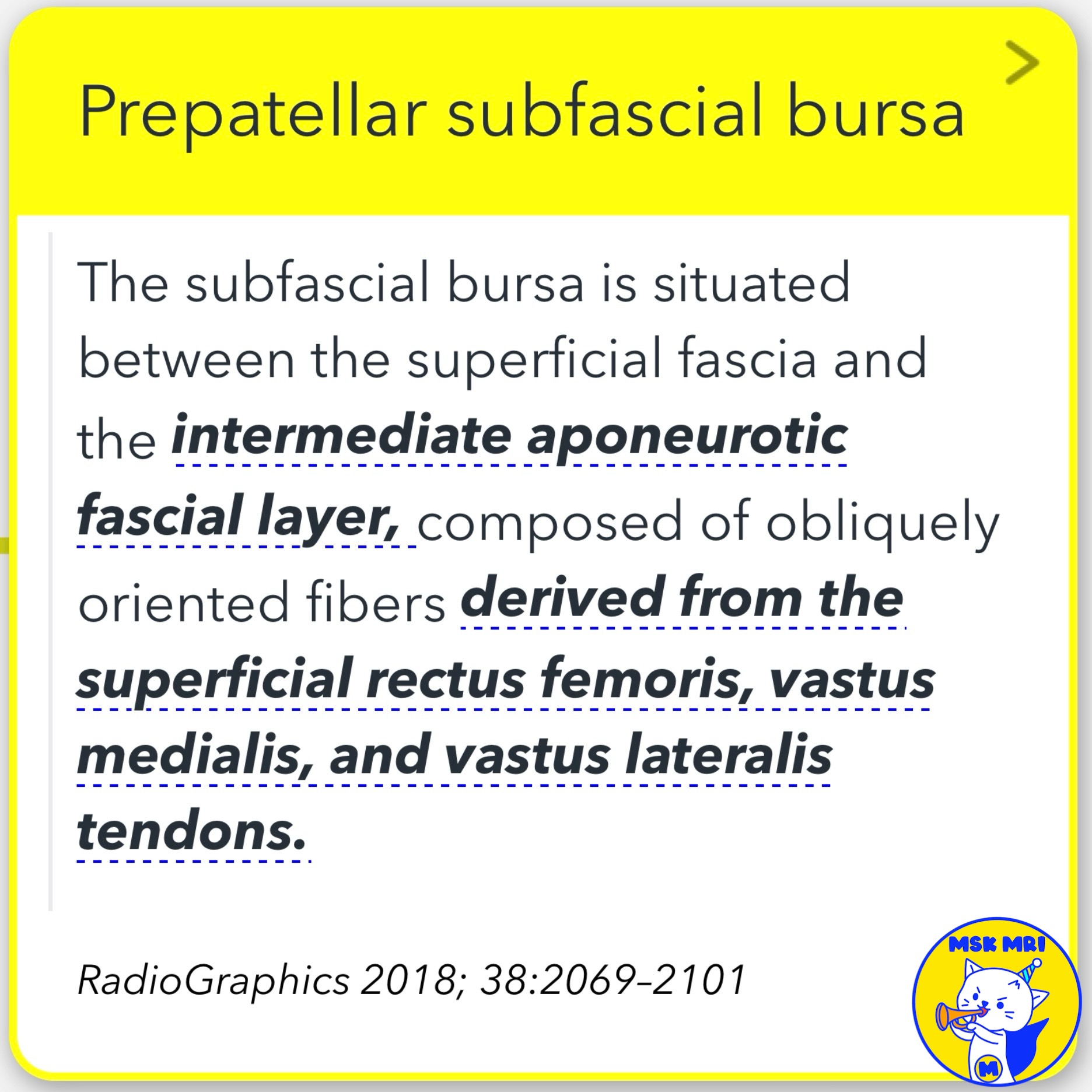
- Prepatellar subfascial bursa
- Intermediate oblique aponeurotic layer
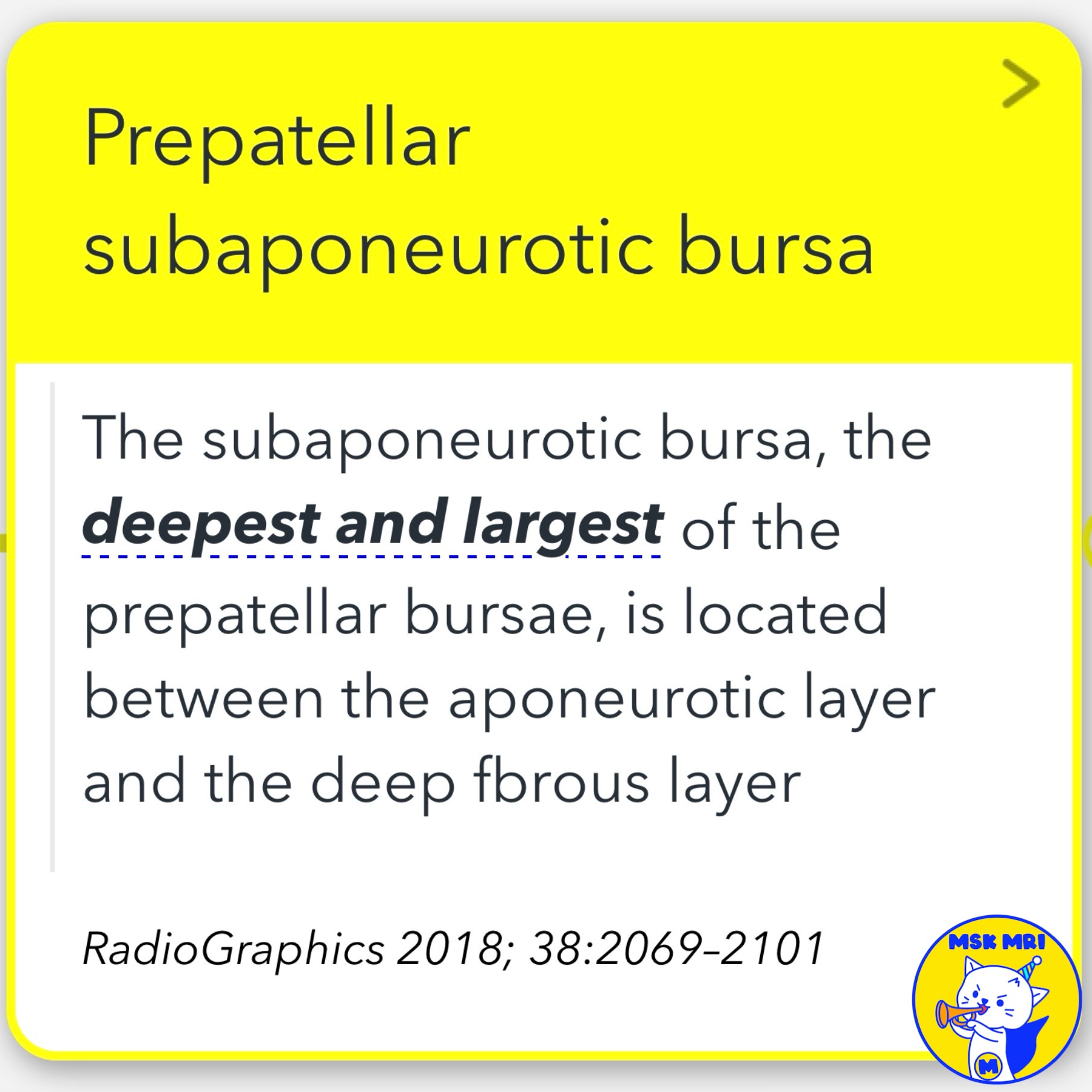
- Prepatellar subaponeurotic bursa
- Prepatellar quadriceps continuation (fibrocartilage)
- Patella bone
✅ Detailed Anatomy
- Prepatellar subcutaneous bursa: Between skin and superficial fascia.
- Prepatellar subfascial bursa: Between superficial fascia and intermediate aponeurotic fascial layer.
- Prepatellar subaponeurotic bursa: Between aponeurotic layer and deep fibrous layer.
- Prepatellar quadriceps continuation: Thickest layer, vertically oriented fibers from rectus femoris tendon adherent to patella.
✅ Imaging Findings
- Superficial bursal fluid: Focal, unilocular fluid arc over patella, patellar tendon, or tibial tubercle.
- Communication between bursae: Unilaminar, bilaminar, or trilaminar appearance when distended.
- Septa within fluid collection: Representing various prepatellar fibrous layers.
References: AJR 2009; 192:W111–W116 RadioGraphics 2018; 38:2069–2101 AJR 2007; 188:W355–W358
"Visualizing MSK Radiology: A Practical Guide to Radiology Mastery"
© 2022 MSK MRI Jee Eun Lee All Rights Reserved.
No unauthorized reproduction, redistribution, or use for AI training.
#prepatellarbursa, #kneeanatomy, #bursa, #patella, #imagingfindings, #trilaminararrangement, #subcutaneousbursa, #subfascialbursa, #subaponeurotibursa, #quadricepscontinuation.


'✅ Knee MRI Mastery > Chap 4BCD. Anterior knee' 카테고리의 다른 글
| (Fig 4-B.11) Quadriceps Tendon Partial Tear (0) | 2024.06.12 |
|---|---|
| (Fig 4-B.10) Prepatellar Bursa Compartmentalization (0) | 2024.06.11 |
| (Fig 4-B.08) Anatomy of Prepatellar Quadriceps Continuation (0) | 2024.06.11 |
| (Fig 4-B.07) Dorsal Defect of the Patella (0) | 2024.06.11 |
| (Fig 4-B.06) Symptomatic Bipartite Patella (1) | 2024.06.10 |