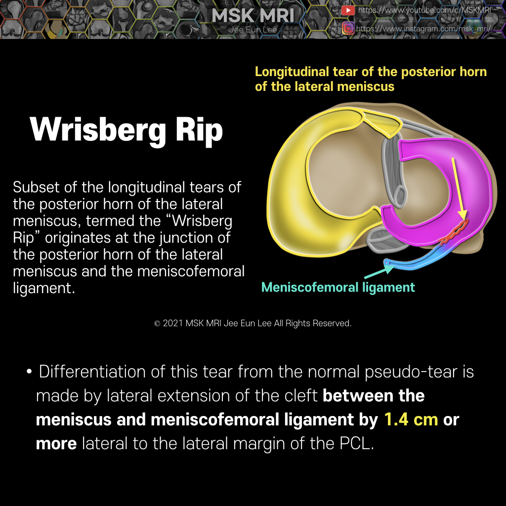
본문 바로가기

Knee MRI/Meniscus62

[Anatomy_19] Anteroinferior popliteomeniscal fascicle 02 The anteroinferior and posterosuperior fascicles were seen on MRI in 97% of patients who had a normal lateral meniscus at arthroscopy Although 45 degree oblique coronal images have been recommended for visualization of the popliteomeniscal fascicles , these structures are often well visualized during the review of routine sagittal acquisitions of the knee The anteroinferior popliteomeniscal fasc.. 2021. 9. 25.
[Anatomy_18] Anteroinferior popliteomeniscal fascicle 01 The anteroinferior popliteomeniscal fascicle (yellow arrow) is seen to course from the inferior aspect of the lateral meniscus posteroinferiorly to blend with fibers of the popliteofibular ligament This fascicle is typically seen on sagittal slices where the fibular head is also visible. In another patient, a component of the anteroinferior popliteomeniscal fascicle which courses directly to the.. 2021. 9. 25.
[Anatomy_17] Tear of posterosuperior Popliteomeniscal fascicle The posterosuperior popliteomeniscal fascicle forms the roof of the popliteus hiatus, extending from the posterosuperior margin of the lateral meniscus to the posterior capsule. The anteroinferior popliteomeniscal fascicle is a coronary ligament, which forms the floor of the popliteus hiatus. As the popliteus tendon passes laterally, it passes beneath the posterosuperior fascicle and above the a.. 2021. 9. 25.
[Shorts #02] Popliteomeniscal fascicle, #shorts Beginning at the lateral aspect of the lateral compartment, the anteroinferior popliteomeniscal fascicle is seen to course from the inferior aspect of the lateral meniscus posteroinferiorly to blend with fibers of the popliteofibular ligament. ​​The posterosuperior popliteomeniscal fascicle is visualized medially to the anteroinferior popliteomeniscal fascicle as the popliteus tendon penetrates .. 2021. 9. 24.
[Anatomy_16] Overview of the popliteomeniscal fascicles The LM has a loose attachment to the joint capsule and to the femur and tibia by means of the meniscal ligaments and is not anchored to the lateral collateral ligament (LCL). Posterior oblique drawing demonstrates posterolateral restraints of the lateral meniscus including the posterosuperior popliteomeniscal fascicle (PSF), anteroinferior popliteomeniscal fascicle (AIF), meniscofemoral ligament.. 2021. 9. 24.
[Anatomy_15] Lateral Meniscus Posterior Root - 03 PRLM has two attachment sites of the intertubercular region and the posterior slope of the lateral tubercle. Contiguous fat-suppressed PD coronal images with a posteroanterior direction demonstrate small, dark signal foci (white arrows) traversing the intertubercular crest and terminating on the medial tubercle, near the tibial attachment of the ACL. These green-yellow arrows show an intercondyl.. 2021. 9. 24.
[Anatomy_14] Lateral Meniscus Posterior Root - 02 PRLM has two attachment sites of intertubercular region and posterior slope of lateral tubercle. This image shows an intercondylar crest insertion component among the two attachment sites. The PRLM inserts mainly in the intertubercular area. It is delineated on MRI as a dark signal line parallel to the tibial plateau on mid-sagittal images with an oblique course and longer length than other root.. 2021. 9. 24.
[Anatomy_13] Lateral Meniscus Posterior Root - 01 The PRLM(lateral meniscus posterior root) provides secondary stability to the anterior cruciate ligament during pivot shift loading. The PRLM has unique features, including its course and configuration. An accurate anatomic understanding of the PRLM is clinically important because this region is difficult to fully visualize arthroscopically. PRLM has two attachment sites of intertubercular regio.. 2021. 9. 24.
[Anatomy_12] Normal Fissured LM Anterior Root The anterior root of lateral meniscus is normally directed obliquely upward on sagittal images and frequently contains increased internal signal intensity. In this location, the increased signal intensity is sometimes referred to as “speckled.” The anterior root of the lateral meniscus differs from the anterior root of the medial meniscus because it often has a prominent fissured appearance simi.. 2021. 9. 24.
[Anatomy_11] Lateral Meniscus Anterior Root Proximity and contiguous fibers between the anterior root ligament of the lateral meniscus (LM) and the anterior cruciate ligament The anterior root of the lateral meniscus attaches anteriorly, along the lateral tibial tubercle, posterolateral to the medial meniscus anterior root. The tibial insertion of the anterior cruciate ligament is found medial to the anterior root Anterior horn transition.. 2021. 9. 24.
[Anatomy_10] Medial Meniscus Posterior Root -02 The posterior root of the medial meniscus was subcategorized into three zones: transition to posterior horn of medial meniscus at the junction between root and posterior horn (a), midsubstance within root ligament proper (b), and entheseal at the tibial attachment point of root ligament The most common location of medial meniscal root ligament tear was junctional, between the ligament and the po.. 2021. 9. 24.
[Anatomy_09] Medial Meniscus Posterior Root -01 The posterior root of the medial meniscus(Red) commences just lateral to the apex of the medial tibial plateau. The yellow line demonstrates where the cartilage of the medial tibial plateau ceases which is another marker for the beginning of the meniscal root. The root attaches along the downward slope of the intercondylar fossa medial and anterior to the posterior cruciate ligament. The medial .. 2021. 9. 24.
[Anatomy_08] Medial Meniscus Anterior Root https://youtu.be/es0xkT3-Wmo The medial meniscus becomes flattened as it transitions into the anterior root medial meniscus (ARMM) This is made up of a single fiber bundle and is the largest of the four roots. The anterior root lateral meniscus (ARLM) and posterior root medial meniscus (PRMM) are made up of multiple fibre bundles(consists of three or more bundles. In 15% of individuals, the ARMM.. 2021. 9. 24.
[Anatomy_07] Attachments of the Meniscus & cruciate ligaments Superior-view drawing shows relations to meniscal roots attachment sites, anterior cruciate ligament, posterior cruciate ligament, transverse ligament, and posterior meniscofemoral Wrisberg's ligament. Anterior root of the medial meniscus: The anterior root of MM inserts along the tibial eminence, anterior to the insertion of the anterior cruciate ligament. Posterior fibers of the anterior horn .. 2021. 9. 24.
[Anatomy_06] Meniscus Vascular Zones and Perimeniscal Plexus https://youtu.be/6Q2lTVFrvPs The meniscus is relatively avascular in adults, except for the peripheral 10% to 25% of the meniscus supplied by the perimeniscal capillary plexus. Notice, on sagittal fat-suppressed PD images, this focal high signal intensity of the peripheral portion of the medial meniscus is not torn, but perimeniscal capillary plexus. The meniscus can be divided into red-red, red.. 2021. 9. 24.
[Anatomy_05] Normal MRI findings of medial stabilizers of the knee https://youtu.be/ZCOejlDr3xw Axial and coronal fat-suppressed proton density-weighted images demonstrate medial collateral bursitis(blue) with an elongated fluid collection interposed between the superficial tibial collateral ligament (yellow) and the meniscofemoral(orange) and meniscotibial(pink) portions of the deep MCL. On axial images, the distended bursa has a characteristic bi- lobulated a.. 2021. 9. 23.
[Anatomy_04] Normal MRI findings of medial stabilizers of the knee https://youtu.be/1PX10ITzBPA The static medial stabilizers include the superficial medial collateral ligament (MCL), the posterior oblique ligament, and the deep capsular (meniscofemoral and meniscotibial) ligaments. Dynamic support is provided by the semimembranosus insertions and the vastus medialis. [Axial image] The MCL has an average thickness of 4.3mm at the femoral attachment and 2.3mm at.. 2021. 9. 23.
[Anatomy_03] Relationship of the static and dynamic structures of the posteromedial corner, https://youtu.be/XLdqNTeHdTc •This figure illustrates the intimate relationship of the static and dynamic structures of the posteromedial corner, including the semimembranosus tendon fascial expansion that attached directly to the posterior horn of the medial meniscus. •TThe medial meniscal posterior meniscocapsular junction is composed of the meniscocapsular ligament superiorly (also known as m.. 2021. 9. 23.
[Anatomy_02] Anatomic relationship of body and posterior horn of medial meniscus and joint capsule https://youtu.be/dnaMoYf8dX8 The anatomic relationship of body and posterior horn of medial meniscus and joint capsule •Axial view illustration of the anatomic relationships of the posterior horn of the medial meniscus (PHMM), posterior capsule, posterior oblique ligament (POL), deep medial collateral ligament (MCL), and semimembranosus tendon. •The attachment of the MM to the joint capsule is m.. 2021. 9. 23.
[Anatomy_01] Overview of normal meniscus anatomy and microstructures https://youtu.be/_lmxTOq_8S8 * MM: The larger, more open C- shaped medial meniscus increases in width from anterior to posterior, resulting in a larger posterior horn compared with the anterior horn when viewed in cross-section. * LM: The more circular lateral meniscus maintains a relatively constant width, resulting in the anterior and posterior horns being nearly equal in size in cross-section.. 2021. 9. 23.