분류 전체보기 727

[Anatomy_13] Ulnotriquetral Ligament, Two Normal Perforation, prestyloid recess, pisotriquetral joint orifice

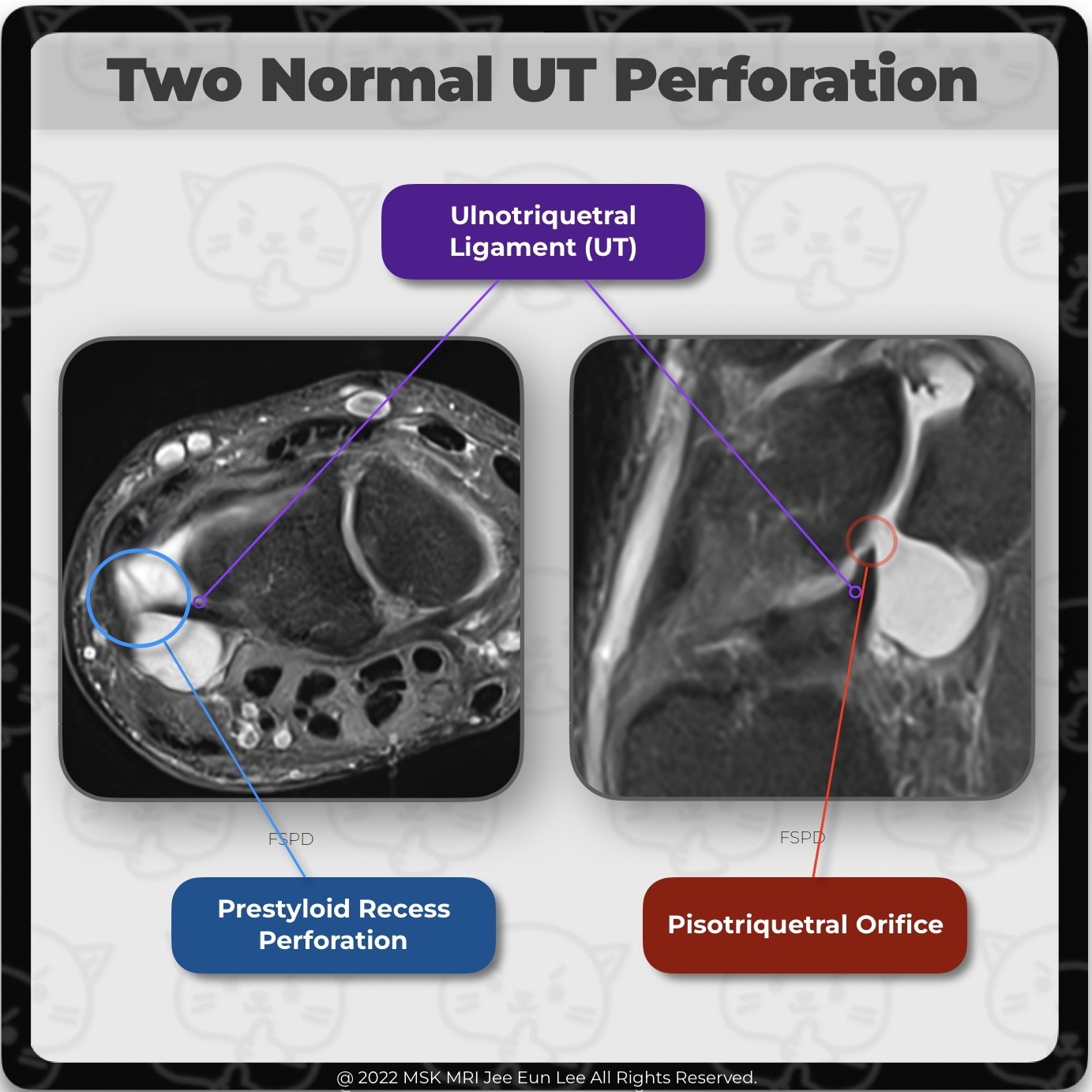
The ulnotriquetral ligament (UT) often has 2 normal perforations: a proximal opening to the prestyloid recess, and a distal pisotriquetral joint orifice. (Left image) The prestyloid recess perforation is found just ulnar to the union between the proximal radioulnar ligament and the UT ligament. (Right image) The pisotriquetral orifice is anterior to the proximal articular surface of the triquetr..

[Anatomy_12] Ulnotriquetral Ligament, TFCC, wrist MRI

The ulnotriquetral ligament (UT) forms the anterior and ulnar part of the ulnocarpal joint capsule together with the ulnolunate ligament. UT shares a common origin and connects the volar radioulnar ligament and thus the triangular fibrocartilage complex to the triquetral bone. #WristMRI, #WristAnatomy, #WristLigament, #WristInstability, #TFCC, #Wrist MRI, #Wrist anatomy, #wrist ligament, #안산에이스병..

[Anatomy_11] Palmar Ulnocarpal Ligaments, TFCC, wrist MRI

IThe ulnocarpal ligament, or ulnocarpal ligamentous complex (UCLC), is anatomically divided into the ulnocapitate, ulnotriquetral, and ulnolunate ligaments. The palmar ulnocarpal ligaments are the superficial ulnocapitate and the deeper ulnolunate and ulnotriquetral ligaments. #WristMRI, #WristAnatomy, #WristLigament, #WristInstability, #TFCC, #Wrist MRI, #Wrist anatomy, #wrist ligament, #안산에이스병..

[Anatomy_10] Radioscapholunate Bundle, Scapholunate Ligament, wrist MRI

MRI shows the normal radioscapholunate bundle attaching to the junction of the volar and proximal segments of the SLL. Scaphoid (S) and lunate (L) bones. Note the low-signal ligamentous appearance of this neurovascular-containing mesocapsule. #WristMRI, #WristAnatomy, #WristLigament, #WristInstability, #Wrist MRI, #Wrist anatomy, #wrist ligament, #안산에이스병원,#Radioscapholunate Bundle, #Scapholunate..

[Anatomy_09] Short radiolunate ligament, Ulnolunate ligament, wrist MRI

The ulnolunate ligament(UL) is continuous with the short radiolunate ligament (SRL) and attaches to the palmar cortex of the lunate just as the short radiolunate ligament does. #WristMRI, #WristAnatomy, #WristLigament, #WristInstability, #Wrist MRI, #Wrist anatomy, #wrist ligament, #안산에이스병원,#short radiolunate ligament, #ulnolunate ligament, #ulnocarpal ligament, #ulnocarpal ligamentous complex,

[Anatomy_08] Scapholunate Ligament Tear, Radioscaphocapitate ligament, Long radiolunate ligament

FSPD image demonstrates full-thickness tear of the membranous portion (not shown) and volar band of the scapholunate ligament (SLL), and partial tear of dorsal band. Note the hyperintense signal and thickening of the radioscaphocapitate (RSC) ligament and long radiolunate ligament (LRL), consistent with grade I injury or sprain. LRL should not be mistaken for intact volar SLL. #WristMRI, #WristA..

[Anatomy_07] Scapholunate Ligament, Long radiolunate ligament, Radioscaphocapitate ligament

The lunate attachment is just dorsal to the long radiolunate ligament (LRL), (=radiolunotriquetral ligament (RLT)) attachment and immediately proximal to the cartilage surface. #WristMRI, #WristAnatomy, #WristLigament, #WristInstability, #Wrist MRI, #Wrist anatomy, #wrist ligament, #안산에이스병원, #Radioscaphocapitate ligament, #Long radiolunate ligament, #Scapholunate Ligament

[Anatomy_06] Interligamentous Sulcus, Radioscaphocapitate ligament, Long radiolunate ligament, Wrist MRI

An interligamentous sulcus (yellow dots), demonstrating fluid signal can sometimes be identified between the radioscaphocapitate (RSC) ligament and long radiolunate ligament (LRL), and should not be mistaken for a tear. #WristMRI, #WristAnatomy, #WristLigament, #WristInstability, #Wrist MRI, #Wrist anatomy, #wrist ligament, #안산에이스병원, #Radioscaphocapitate ligament, #Long radiolunate ligament, #In..

[Anatomy_05] Radioscaphocapitate ligament, Long radiolunate ligament, Short radiolunate ligament

At the level of the scaphoid (left), the radioscaphocapitate (RSC) ligament is seen palmar to the waist of the scaphoid, being distal to the long radiolunate ligament (LRL). At the level of the lunate (right), MRI shows a short radiolunate ligament (SRL) passing from the volar lip of the distal radius at the lunate facet, to the volar lunate cortex. Immediately distal to the SRL is the LRL, foll..

[Anatomy_04] Ganglion cyst, Long radiolunate ligament, Radioscaphocapitate ligament, Wrist MRI

Ganglion cysts are also commonly associated with extrinsic ligaments. The volar ganglion cyst with a loculated component interdigitating with the fibers of the long radiolunate (LRL) or radiolunotriquetral ligament (RLT). #WristMRI, #WristAnatomy, #WristLigament, #WristInstability, #Wrist MRI, #Wrist anatomy, #wrist ligament, #안산에이스병원, #Radioscaphocapitate ligament, #Long radiolunate ligament, #..