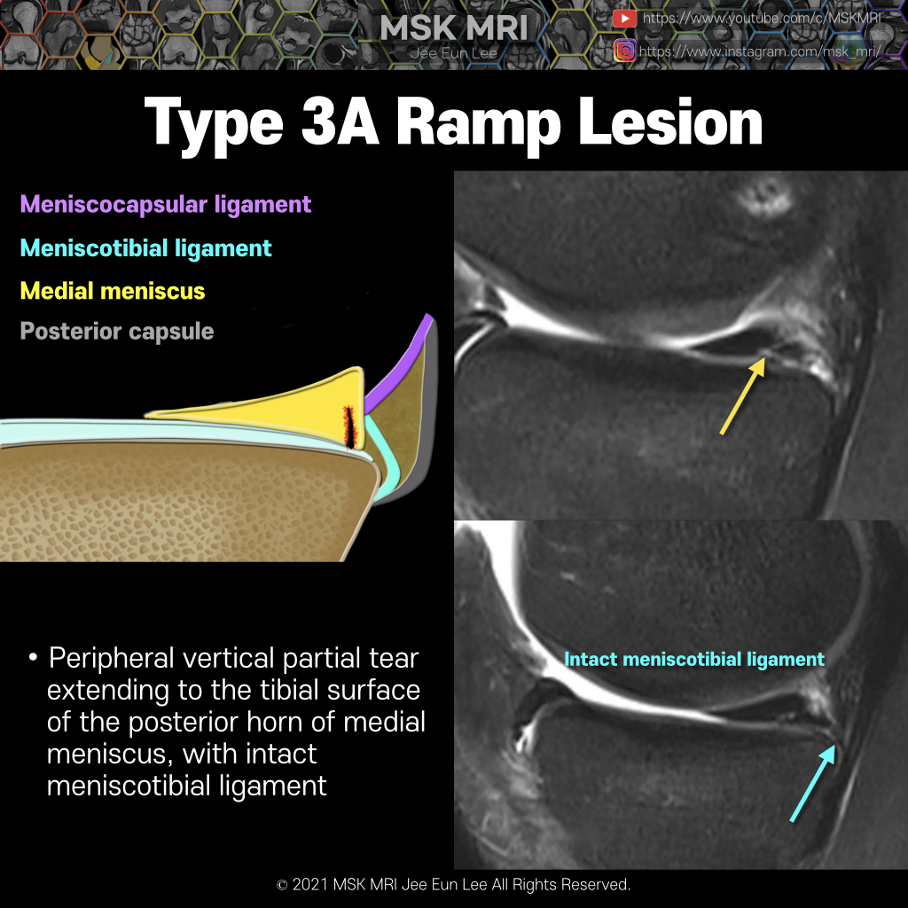
본문 바로가기

ACL injury26

[Tear_12] Wrisberg Rip_Longitudinal-Vertical Tears -10 Wrisberg rip, also known as zip tear or zipper tear is the longitudinal tear of the posterior horn of the lateral meniscus. Wrisberg rip occurs at the junction of the meniscofemoral ligament and the posterior horn of the lateral meniscus. Axial oblique reformatted image demonstrates the course of the ligament of Humphrey anterior to the PCL (blue arrows) as it joins to the meniscal fragment peri.. 2021. 10. 10.
[Tear_11] Wrisberg Rip_Longitudinal-Vertical Tears -09 Posterior oblique drawing demonstrates PCL, LM, meniscofemoral ligaments (Humphry ligament and Wrisberg ligament) The meniscofemoral ligaments originate from the posterior horn of the lateral meniscus and insert onto the lateral aspect of the posterior medial femoral condyle, with the Humphry ligament anterior to the posterior cruciate ligament (PCL) and the Wrisberg ligament posteriorly Wrisber.. 2021. 10. 10.
[Tear_10] Longitudinal-Vertical Tears_ ACL tear -08 There is a close association between ACL tears and peripheral longitudinal tears in the posterior horns of both the medial and lateral menisci. Acute ACL injury is associated with more lateral meniscal tears, while chronic ACL deficiency is associated with more medial meniscal tears,many of which require meniscectomy. So early ACL reconstruction is recommended also for the prevention of secondar.. 2021. 10. 10.
[Tear_09] Wrisberg type discoid lateral meniscus -07 Wrisberg-variant discoid lateral meniscus can be interpreted as peripheral longitudinal tear or popliteomeniscal fascicles tear. Watanabe classification recognizes three distinct variants of discoid meniscus, complete variant, partial variant, and Wrisberg variant. The Wrisberg-variant discoid lateral meniscus is an unstable type that lacks posterior ligament attachments, having only an attachme.. 2021. 10. 9.
[Tear_08] Tear of popliteomeniscal fascicles -06 It is sometimes difficult to identify peripheral longitudinal tears in the posterior horn of the lateral meniscus because of the complex anatomy and posterior attachments of the capsule and popliteomeniscal fascicles. Far peripheral longitudinal tears can be more difficult to detect With an “extra” vertical line or layer in the meniscal periphery, it can be a subtle finding that indicates a tear.. 2021. 10. 9.