본문 바로가기

Meniscus29

(Fig 1-A.18) anterior meniscofemoral ligament of the medial meniscus, infrapatellar plica 
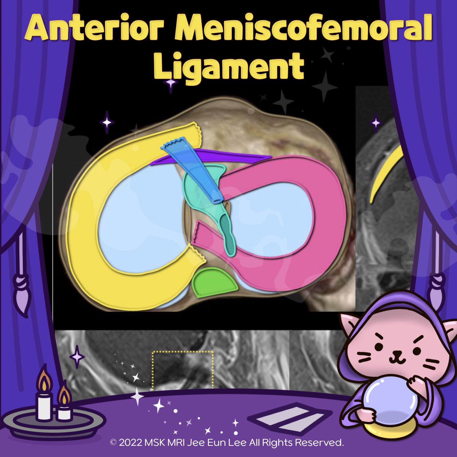
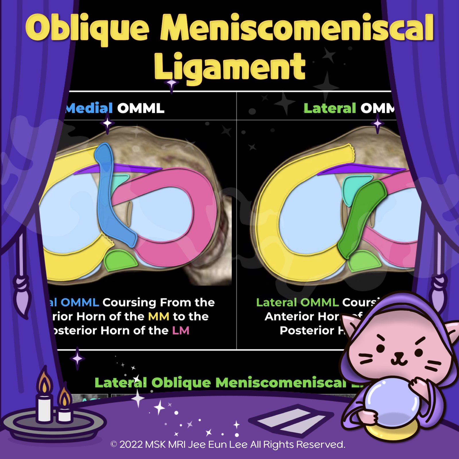
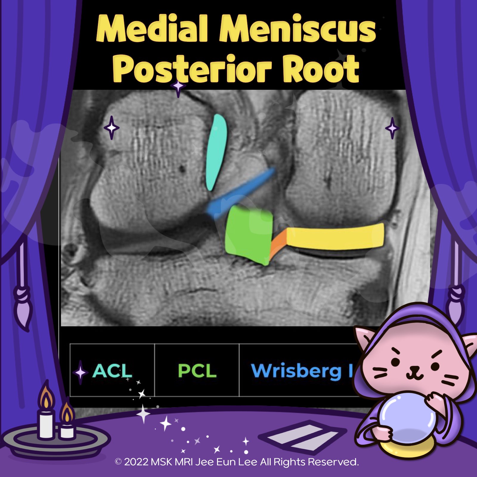
https://youtu.be/mxyxCuCKwzM https://youtu.be/kEuMzUrHD0M https://youtu.be/kEuMzUrHD0M"Anomalous insertion of the medial meniscus (AIMM) into the anterior cruciate ligament (ACL) is an uncommon condition, seen in about 0.52% of arthroscopy cases. It involves a ligament extending to the ACL from the anterior horn of the medial meniscus, near the root. This variant accessory insertion appears on s.. 2024. 1. 16.
(Fig 1-A.17) Oblique meniscomeniscal ligament https://youtu.be/fyX1V1RrctQ https://youtu.be/kEuMzUrHD0M Aspect Details Ligament Type Oblique Meniscomeniscal Ligaments (Intermeniscal Ligament) Prevalence 1% to 4% Location Transverse the intercondylar notch between the anterior and posterior cruciate ligaments Naming Based on anterior attachment sites Medial OMML Originates from the anterior horn of the medial meniscus extends through the int.. 2024. 1. 15.
(Fig 1-A.15) Additional peripheral attachments to the MM https://youtu.be/C-I1T1POols https://youtu.be/kEuMzUrHD0M The knee joint capsule, part of layer III, forms the deep MCL, with its meniscotibial and meniscofemoral components, along the medial aspect of the knee. It then continues posteriorly, where it is reinforced externally by the POL and expansions from the semimembranosus. More posteriorly, the posteromedial joint capsule passes deep to the .. 2024. 1. 13.
(Fig 1-A.06) Medial Meniscus Posterior Root, Anatomy, degeneration, tear https://youtu.be/CjYENTgOT3Y https://youtu.be/kEuMzUrHD0M "🧐 Deep Dive into the Meniscofemoral Ligament (MFL) Anatomy! ✨ The MFL, a critical component of the knee, originates from the posterior horn of the lateral meniscus and inserts onto the lateral aspect of the posterior medial femoral condyle. It plays a key role in the stability and function of the knee joint. 🔍 Let's break it down: 1️⃣ Th.. 2024. 1. 7.
(Fig 1-A.05) Anatomy of the Lateral Meniscus Posterior Root, two components, intertubercular area https://youtu.be/-lNJo4zWnqs https://youtu.be/kEuMzUrHD0M PRLM has two attachment sites of the posterior slope of the lateral tubercle (1st coronal image) and intertubercular region (other coronal images). - major component: intertubercular area, anterior extension into medial tubercle along the intertubercular crest, just posteromedial to tibial attachment of anterior cruciate ligament (; ACL) .. 2024. 1. 6.
(Fig 1-A.06) Medial Meniscus Posterior Root, Anatomy, degeneration, tear https://youtu.be/GvR9ULGrDOw https://youtu.be/kEuMzUrHD0M Category Description Insertion Points - Inserts along the downward slope of the intercondylar fossa, anterior to the posterior cruciate ligament. - Extends from the tibial attachment point to just lateral from the articular cartilage inflection point of the medial tibial plateau. - Commences just lateral to the apex of the medial tibial p.. 2024. 1. 5.
[HYPERLINK] Meniscus Anatomy, root, Discoid meniscus, Wrisberg ligament, Poplite Meniscus Anatomy, root, Discoid meniscus, Wrisberg ligament, Popliteomeniscal fascicles, Variants and Pitfalls ​ Anatomy ​ [Meniscus Anatomy_01] Knee MRI, Anatomy, anterior root, posterior root, medial lateral meniscus [Meniscus Anatomy_02] Popliteomeniscal fascicles (anteroinferior, posterosuperior ) [Meniscus Anatomy_03] Humphry and Wrisberg ligaments, meniscotibial ligament, knee MRI [Meniscu.. 2022. 5. 14.
[HYPERLINK] Knee MRI checklist, meniscus, tear, ACL, PCL, MCL, LCL, Extensor mec Knee MRI checklist ​ [Knee_01] MRI check list, meniscus, anatomy, root, horizontal tear, vertical tear, meniscus tear [Knee_02] Meniscus tear, radial tear, root tear, flap, parrot beak , Bucket handle tear, KNEE MRI [Knee_03] meniscectomy, meniscus repair, Transtibial pullout repair, transplantation, KNEE MRI [Knee_04] ACL, PCL, Tear, Reconstruction, femoral tunnel, tibial tunnel, impingement, g.. 2022. 5. 14.
[Tear_08] Tear of popliteomeniscal fascicles -06 It is sometimes difficult to identify peripheral longitudinal tears in the posterior horn of the lateral meniscus because of the complex anatomy and posterior attachments of the capsule and popliteomeniscal fascicles. Far peripheral longitudinal tears can be more difficult to detect With an “extra” vertical line or layer in the meniscal periphery, it can be a subtle finding that indicates a tear.. 2021. 10. 9.
[Tear_07] Far Peripheral Longitudinal Meniscal Tear -05 On the left image, we can easily see the longitudinal tear, but on the right image it is very difficult to identify peripheral longitudinal tear. Like this, longitudinal tears that occur further peripherally can be more difficult to detect. When the tear extends through the meniscal substance it is readily identified against the contrast afforded by the low signal meniscal tissue. or, Identifica.. 2021. 10. 9.
[Tear_06] Far Peripheral Tear Longitudinal Meniscal Tear -04 Here is a full-thickness peripheral longitudinal tear of the posterior horn of lateral meniscus. There is an extra vertical line or layer in the periphery of lateral meniscus, indicative of far peripheral longitudinal tear. The tear is parallel and adjacent to the popliteus tendon. It is sometimes difficult to identify peripheral longitudinal tears in the posterior horn of the lateral meniscus b.. 2021. 10. 9.
[Tear_05] Inferior Surface Longitudinal Meniscal Tear -03 Longitudinal tears are diagnosed on MRI by the presence of a vertical line of increased signal intensity contacting the superior, inferior, or both surfaces of the meniscus. The longitudinal tear extends to the inferior meniscal surface and courses parallel to the long axis of the meniscus on corresponding PD FSE axial image. [Tear_04] case demonstrates vertically oriented increased signal inten.. 2021. 10. 9.
[Tear_04] Complete Longitudinal Meniscal Tear -02 Longitudinal tears almost always involve the posterior horn in both the medial and lateral menisci. They are diagnosed on MRI by the presence of a vertical line of increased signal intensity contacting the superior, inferior, or both surfaces of the meniscus. MR images showing vertically oriented increased signal intensity contacting both articular surfaces of the body and posterior horn of the .. 2021. 10. 9.
[Tear_03] Longitudinal Meniscal Tear -01 Left drawing shows a typical longitudinal tear that divides the meniscus into inner and outer halves, and the right drawing shows the expected appearance of the tear. Longitudinal tear with peripheral vertical orientation through the circumferential collagen fibers. Longitudinal tears run perpendicular to the tibial plateau and parallel to the long axis of the meniscus. Unlike horizontal or radi.. 2021. 10. 9.
[Shorts #02] Popliteomeniscal fascicle, #shorts Beginning at the lateral aspect of the lateral compartment, the anteroinferior popliteomeniscal fascicle is seen to course from the inferior aspect of the lateral meniscus posteroinferiorly to blend with fibers of the popliteofibular ligament. ​​The posterosuperior popliteomeniscal fascicle is visualized medially to the anteroinferior popliteomeniscal fascicle as the popliteus tendon penetrates .. 2021. 9. 24.
[Anatomy_16] Overview of the popliteomeniscal fascicles The LM has a loose attachment to the joint capsule and to the femur and tibia by means of the meniscal ligaments and is not anchored to the lateral collateral ligament (LCL). Posterior oblique drawing demonstrates posterolateral restraints of the lateral meniscus including the posterosuperior popliteomeniscal fascicle (PSF), anteroinferior popliteomeniscal fascicle (AIF), meniscofemoral ligament.. 2021. 9. 24.
[Anatomy_15] Lateral Meniscus Posterior Root - 03 PRLM has two attachment sites of the intertubercular region and the posterior slope of the lateral tubercle. Contiguous fat-suppressed PD coronal images with a posteroanterior direction demonstrate small, dark signal foci (white arrows) traversing the intertubercular crest and terminating on the medial tubercle, near the tibial attachment of the ACL. These green-yellow arrows show an intercondyl.. 2021. 9. 24.
[Anatomy_14] Lateral Meniscus Posterior Root - 02 PRLM has two attachment sites of intertubercular region and posterior slope of lateral tubercle. This image shows an intercondylar crest insertion component among the two attachment sites. The PRLM inserts mainly in the intertubercular area. It is delineated on MRI as a dark signal line parallel to the tibial plateau on mid-sagittal images with an oblique course and longer length than other root.. 2021. 9. 24.
[Anatomy_13] Lateral Meniscus Posterior Root - 01 The PRLM(lateral meniscus posterior root) provides secondary stability to the anterior cruciate ligament during pivot shift loading. The PRLM has unique features, including its course and configuration. An accurate anatomic understanding of the PRLM is clinically important because this region is difficult to fully visualize arthroscopically. PRLM has two attachment sites of intertubercular regio.. 2021. 9. 24.
[Anatomy_12] Normal Fissured LM Anterior Root The anterior root of lateral meniscus is normally directed obliquely upward on sagittal images and frequently contains increased internal signal intensity. In this location, the increased signal intensity is sometimes referred to as “speckled.” The anterior root of the lateral meniscus differs from the anterior root of the medial meniscus because it often has a prominent fissured appearance simi.. 2021. 9. 24.
[Anatomy_11] Lateral Meniscus Anterior Root Proximity and contiguous fibers between the anterior root ligament of the lateral meniscus (LM) and the anterior cruciate ligament The anterior root of the lateral meniscus attaches anteriorly, along the lateral tibial tubercle, posterolateral to the medial meniscus anterior root. The tibial insertion of the anterior cruciate ligament is found medial to the anterior root Anterior horn transition.. 2021. 9. 24.