"🧐 Deep Dive into the Meniscofemoral Ligament (MFL) Anatomy!
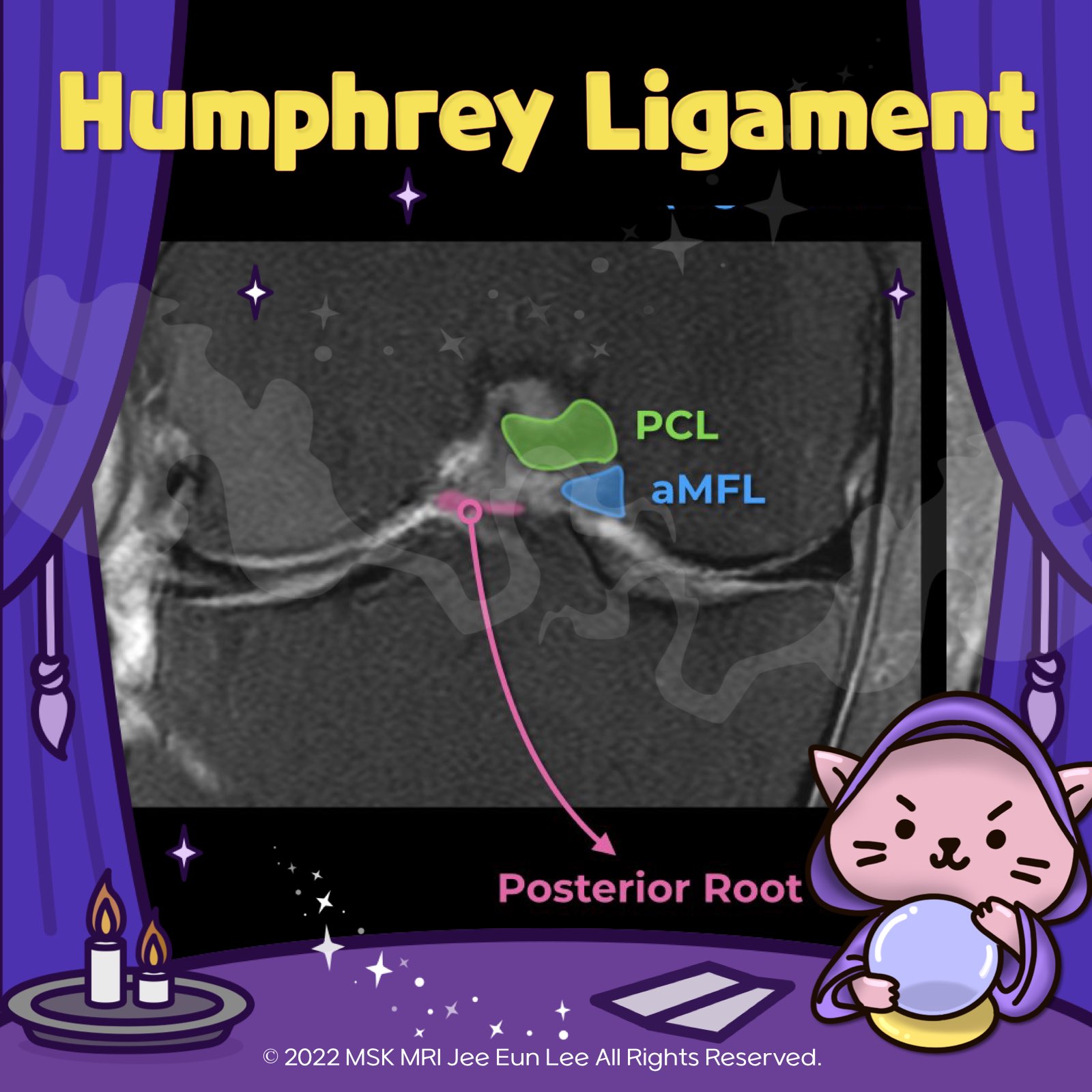
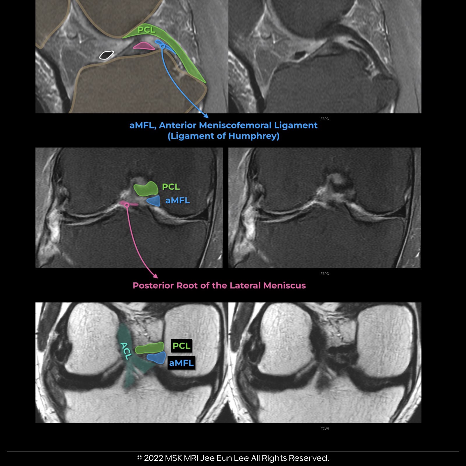
✨ The MFL, a critical component of the knee, originates from the posterior horn of the lateral meniscus and inserts onto the lateral aspect of the posterior medial femoral condyle. It plays a key role in the stability and function of the knee joint.
🔍 Let's break it down:
1️⃣ The aMFL (anterior MFL), also known as the Ligament of Humphrey, originates on the Medial Femoral Condyle and is typically found distal to the PMB of the PCL.
2️⃣ The pMFL (posterior MFL), or the Ligament of Wrisberg, is most commonly observed just proximal to the PM Bundle Attachment of the PCL.
#MeniscofemoralLigament #MFL #KneeAnatomy #LigamentOfHumphrey #LigamentOfWrisberg #aMFL #pMFL #PCL #MedialFemoralCondyle #Orthopedics
© 2022 MSK MRI Jee Eun Lee All Rights Reserved.
You may not distribute or commercially exploit the content.
Nor may you transmit it or store it on any other website or other forms of the electronic retrieval system.
If you would like to use an image or video for anything other than personal use, please contact me. (jamaisvu1977@gmail.com), (jamaisvu77@naver.com) or instagram (msk_mri)