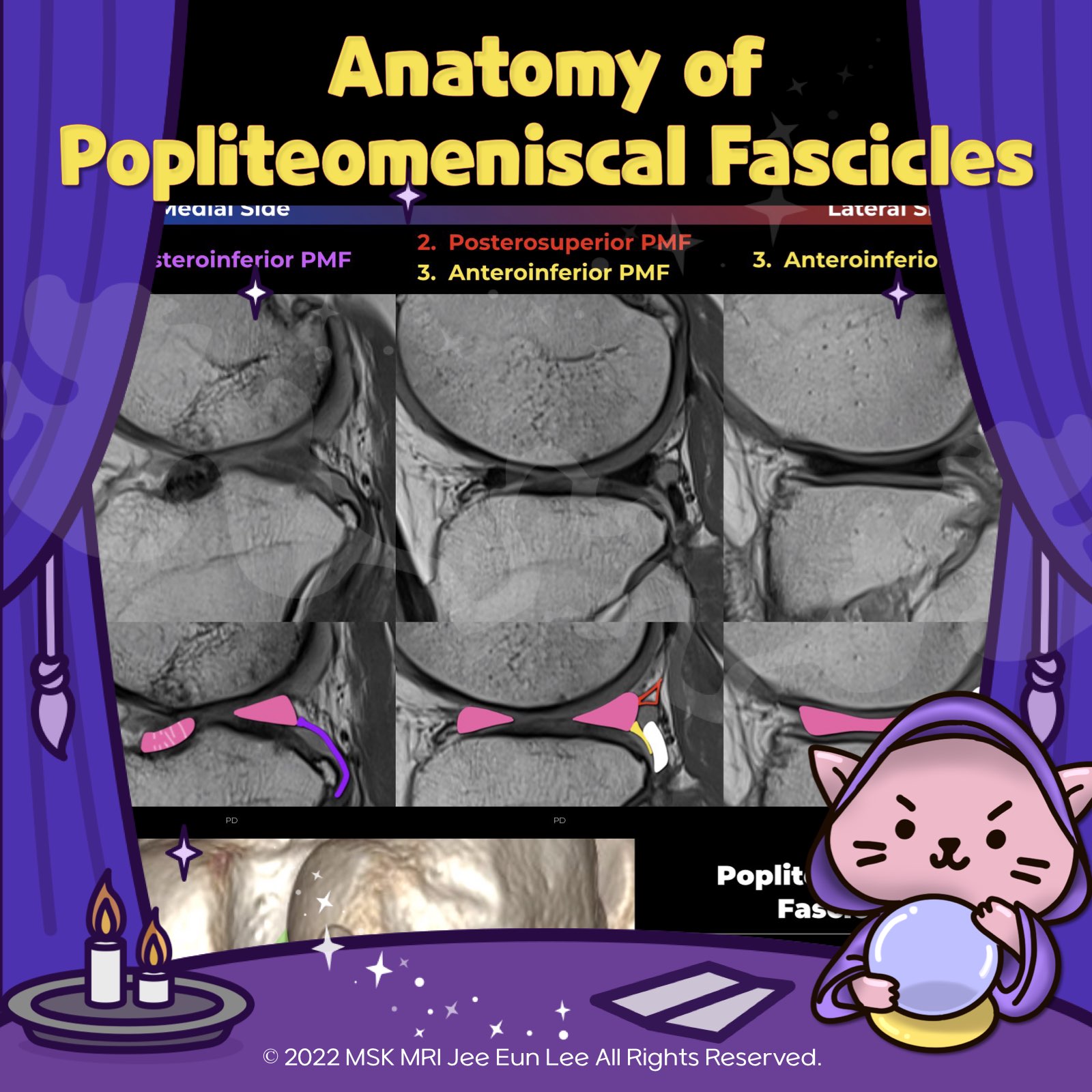
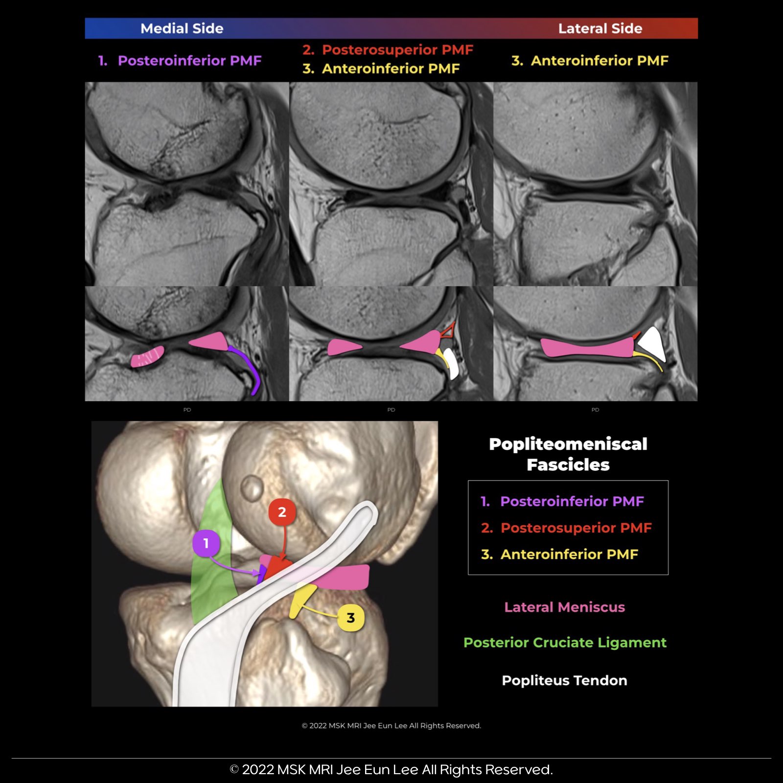
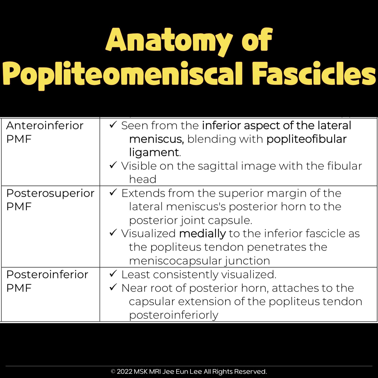
| Fascicle | Location and Appearance |
| Anteroinferior Popliteomeniscal Fascicle |
Visible on sagittal slices near the fibular head; extends posteroinferiorly from the lateral side of the lateral meniscus (LM) and merges with the popliteus tendon; conjoined attachment with the popliteofibular ligament at the fibula’s styloid process; variable appearance. |
| Posterosuperior Popliteomeniscal Fascicle |
Extends from the superior margin of the LM's peripheral posterior horn to the posterior joint capsule, above the popliteus tendon; visualized medial to the anteroinferior fascicle, especially as the popliteus tendon penetrates the meniscocapsular junction. |
| Posteroinferior Popliteomeniscal Fascicle |
Least consistently visualized; near the posterior horn's root; extends from the inferior margin of the LM posteroinferiorly to a capsular extension of the popliteus tendon; in cross-section, resembles a torn inferior flap of the meniscus. |
© 2022 MSK MRI Jee Eun Lee All Rights Reserved. You may not distribute or commercially exploit the content. Nor may you transmit it or store it on any other website or other forms of the electronic retrieval system. If you would like to use an image or video for anything other than personal use, please contact me. (jamaisvu1977@gmail.com), (jamaisvu77@naver.com) or instagram (msk_mri)
'✅ Knee MRI Mastery > Chap 1. Meniscus' 카테고리의 다른 글
| (Fig 1-A.14) anatomy of the Posterior Medial Capsule (PMC) (0) | 2024.01.12 |
|---|---|
| (Fig 1-A.13) normal popliteomeniscal fascicle deficiency (2) | 2024.01.11 |
| (Fig 1-A.11) Meniscofemoral ligaments (Humphrey ligament) (0) | 2024.01.09 |
| (Fig 1-A.10) Meniscofemoral ligament (Wrisberg ligament) (0) | 2024.01.07 |
| (Fig 1-A.06) Medial Meniscus Posterior Root, Anatomy, degeneration, tear (0) | 2024.01.07 |