Click the link to purchase on Amazon 🎉📚
==============================================
🎥 Check Out All Videos at Once! 📺
👉 Visit Visualizing MSK Blog to explore a wide range of videos! 🩻
https://visualizingmsk.blogspot.com/?view=magazine
📚 You can also find them on MSK MRI Blog and Naver Blog! 📖
https://www.instagram.com/msk_mri/
Click now to stay updated with the latest content! 🔍✨
==============================================
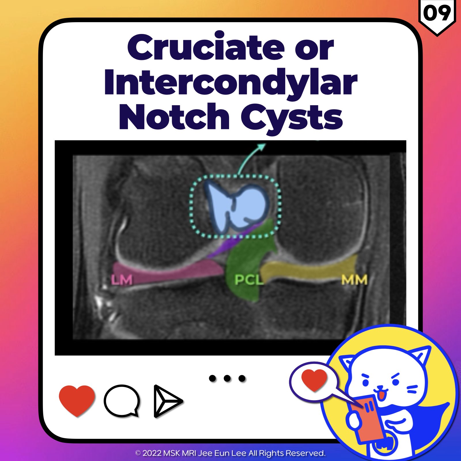
★ Cruciate or Intercondylar Notch Cysts ★
1️⃣ Definition:
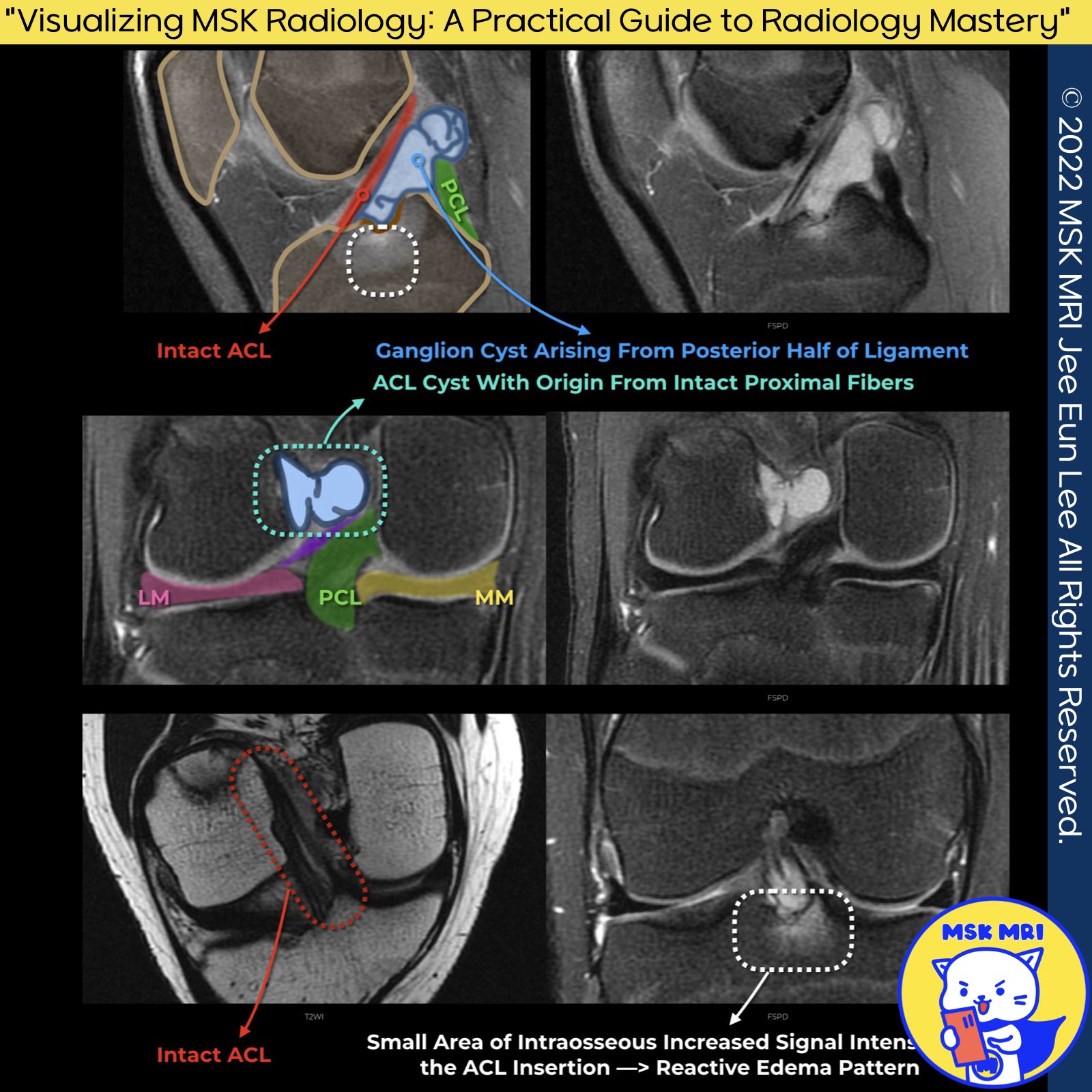
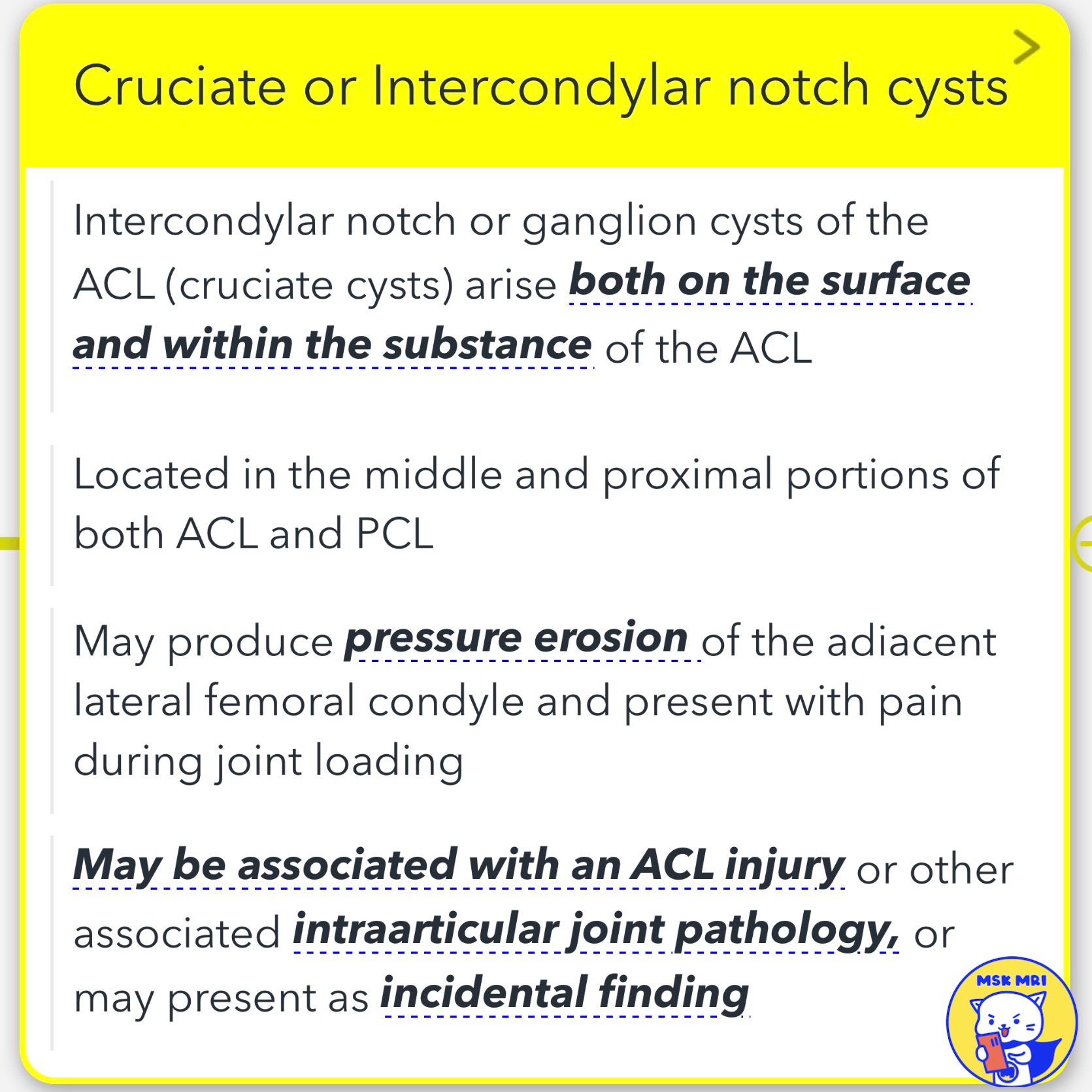
Intercondylar notch or ganglion cysts of the ACL, also known as cruciate cysts, are pathological formations that can arise both on the surface and within the Anterior Cruciate Ligament (ACL) substance.
2️⃣ Location:
These cysts are typically found in the middle and proximal portions of both the ACL and the Posterior Cruciate Ligament (PCL), highlighting their potential to affect critical structures within the knee joint.
3️⃣ Clinical Implications:
📌 Pressure Erosion: The cysts may produce pressure erosion of the adjacent lateral femoral condyle, a condition that could be symptomatic and present with pain, especially during joint loading activities.
4️⃣ Association with Injuries:
A potential association between these cysts and ACL injuries and other intraarticular joint pathologies indicates a need for careful diagnostic evaluation.
5️⃣ Incidental Findings:
In some cases, cruciate or intercondylar notch cysts may present as incidental findings during imaging for unrelated conditions, suggesting a varied clinical significance.
"Visualizing MSK Radiology: A Practical Guide to Radiology Mastery"
© 2022 MSK MRI Jee Eun Lee All Rights Reserved.
#VisualizingMSK #aclanatomy #ACL #ACLcyst #cruciatecyst #aclinjury
You may not distribute or commercially exploit the content. Nor may you transmit it or store it on any other website or other forms of the electronic retrieval system.
'✅ Knee MRI Mastery > Chap 2.ACL and PCL' 카테고리의 다른 글
| (Fig 2-A.11) Complete tear versus mucoid degeneration of the ACL. (0) | 2024.02.17 |
|---|---|
| (Fig 2-A.10) Mucinous or mucoid degeneration of the ACL (0) | 2024.02.16 |
| (Fig 2-A.07) Normal ACL on oblique coronal images (0) | 2024.02.16 |
| (Fig 2-A.06) Normal ACL on axial images (0) | 2024.02.16 |
| (Fig 2-A.04) Normal ACL on coronal images, anteromedial and posterolateral bundles of ACL (0) | 2024.02.16 |
