Click the link to purchase on Amazon 🎉📚
==============================================
🎥 Check Out All Videos at Once! 📺
👉 Visit Visualizing MSK Blog to explore a wide range of videos! 🩻
https://visualizingmsk.blogspot.com/?view=magazine
📚 You can also find them on MSK MRI Blog and Naver Blog! 📖
https://www.instagram.com/msk_mri/
Click now to stay updated with the latest content! 🔍✨
==============================================
1️⃣ Single vs. Two-Stage Surgery
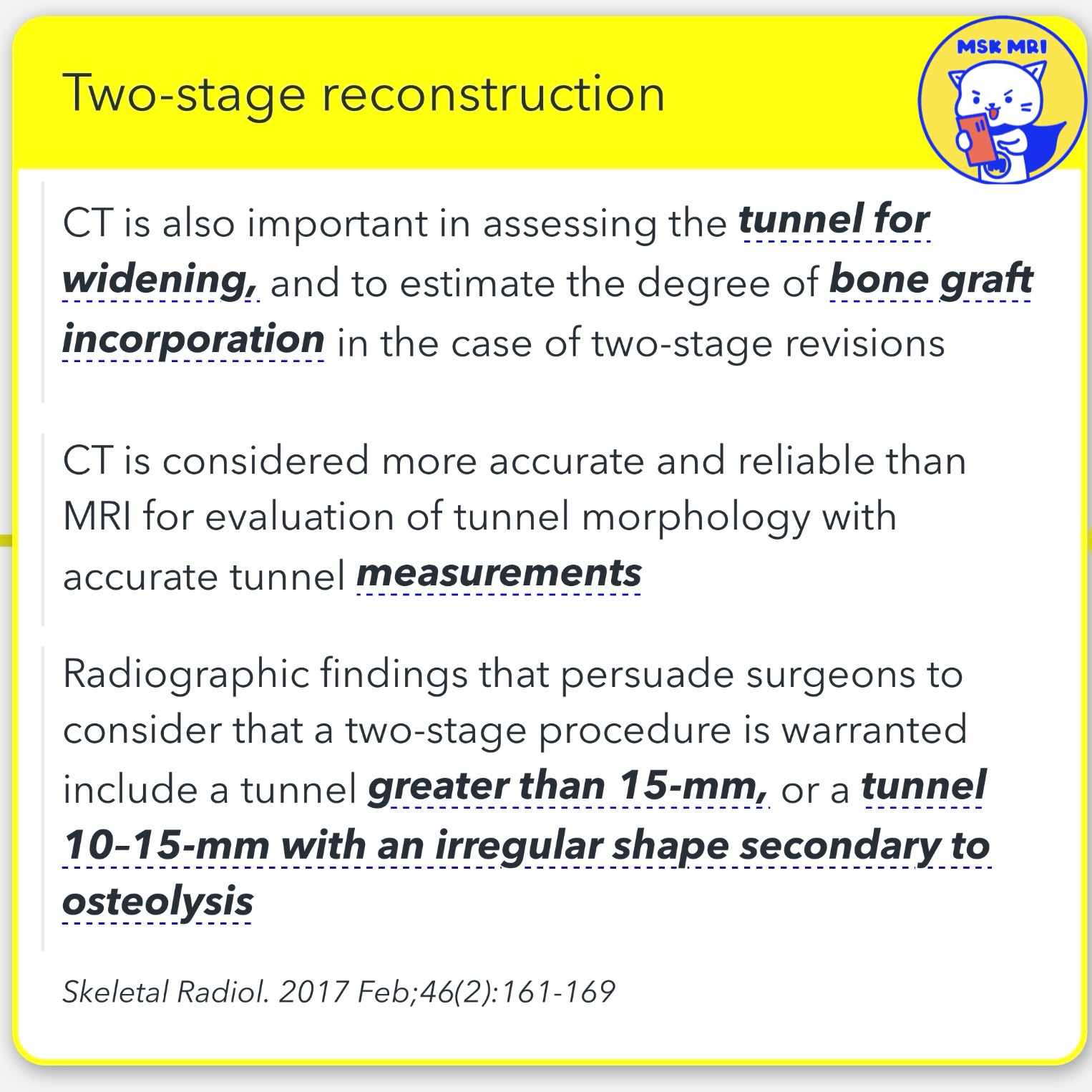
- Studies published to date support that a tunnel diameter greater than 15 mm will require two-stage surgery when the original tunnels are in anatomical position.
- Conversely, a revision with a tunnel diameter of less than 10 mm can typically be accomplished in a single surgery. For tunnels measuring 10–15 mm, the approach differs based on the tunnel shape, position, and the treating surgeon’s preference.
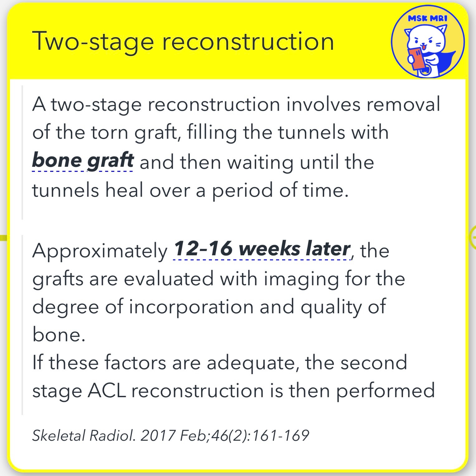
2️⃣ Two-Stage Reconstruction Explained
- Initial Stage: Removal and Filling
A two-stage reconstruction involves removing the torn graft and filling the tunnels with bone graft. - This is then left to heal over some time, typically 12–16 weeks.
- Second Stage: Evaluation and Reconstruction
- Approximately 12–16 weeks later, the grafts are evaluated with imaging to assess the degree of incorporation and the quality of the bone.
- If these factors are deemed adequate, the second stage of the ACL reconstruction is then performed.
3️⃣ Importance of CT in Two-Stage Reconstructions
- CT scans play a crucial role in assessing the tunnel for widening and estimating the degree of bone graft incorporation in cases of two-stage revisions.
- It is considered more accurate and reliable than MRI for evaluating tunnel morphology with precise tunnel measurements.
- Skeletal Radiol. 2017 Feb;46(2):161-169
https://visualizingmsk.blogspot.com/?view=magazine
Visualizing MSK Radiology
visualizingmsk.blogspot.com
"Visualizing MSK Radiology: A Practical Guide to Radiology Mastery"
© 2022 MSK MRI Jee Eun Lee All Rights Reserved.
#VisualizingMSK #ACLinjuries #KneeMRI #ACLtear #ACLReconstruction #BoneGraftHealing #TwoStageSurgery
You should not distribute or commercially exploit the content.
You should not transmit or store it on any other website or electronic retrieval system.


