==============================================
⬇️✨⬇️🎉⬇️🔥⬇️📚⬇️
Click the link to purchase on Amazon 🎉📚
==============================================
🎥 Check Out All Videos at Once! 📺
👉 Visit Visualizing MSK Blog to explore a wide range of videos! 🩻
https://visualizingmsk.blogspot.com/?view=magazine
📚 You can also find them on MSK MRI Blog and Naver Blog! 📖
https://www.instagram.com/msk_mri/
Click now to stay updated with the latest content! 🔍✨
==============================================
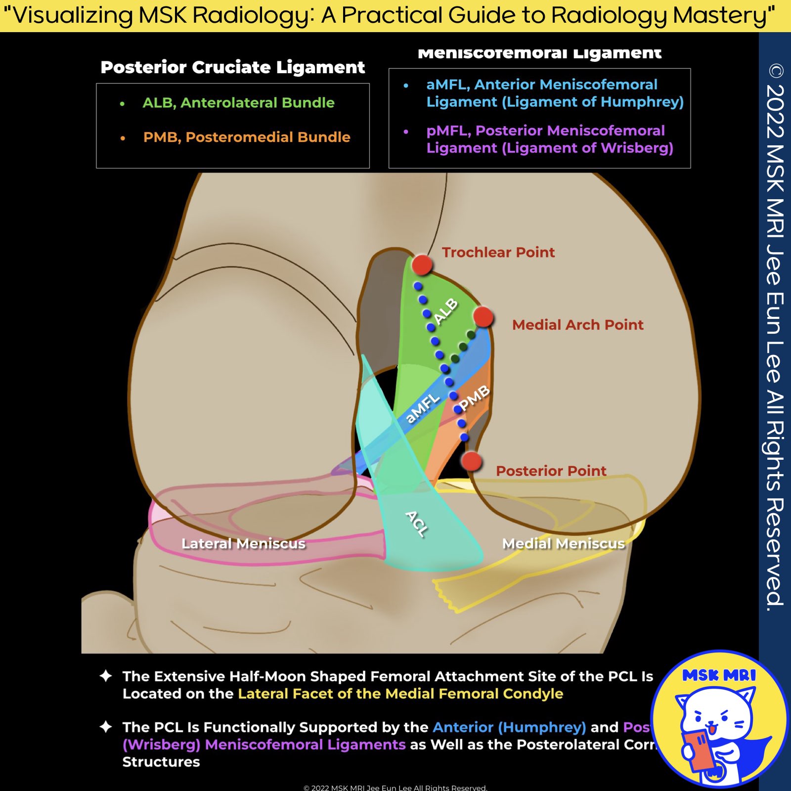
1️⃣ Femoral Attachment of the PCL
- Location: The femoral attachment of the PCL is situated on the lateral facet of the medial femoral condyle.
This extensive half-moon shaped site encompasses the roof of the intercondylar notch anteriorly, is bound proximally by the medial intercondylar ridge, and reaches the margin of the articular cartilage of the medial femoral condyle distally. - Source: Knee Surg Sports Traumatol Arthrosc. 2021 Mar;29(3):672-681.
2️⃣ Femoral Bony Landmarks
- Variability and Consistency: The femoral attachments are notable for variability, contrasting the relatively consistent size and shape of the tibial insertion.
- Identification: The medial intercondylar ridge marks the superior border of the AL and PM bundle origins, with the medial bifurcate ridge separating the two bundles.
- Source: J Knee Surg. 2021 Apr;34(5):499-508.
3️⃣ Modified Quadrant Method
- Methodology: Utilizing a modified quadrant method on strict lateral radiographs of the medial femoral condyle:
- Source: Knee Surg Sports Traumatol Arthrosc. 2021 Mar;29(3):672-681.
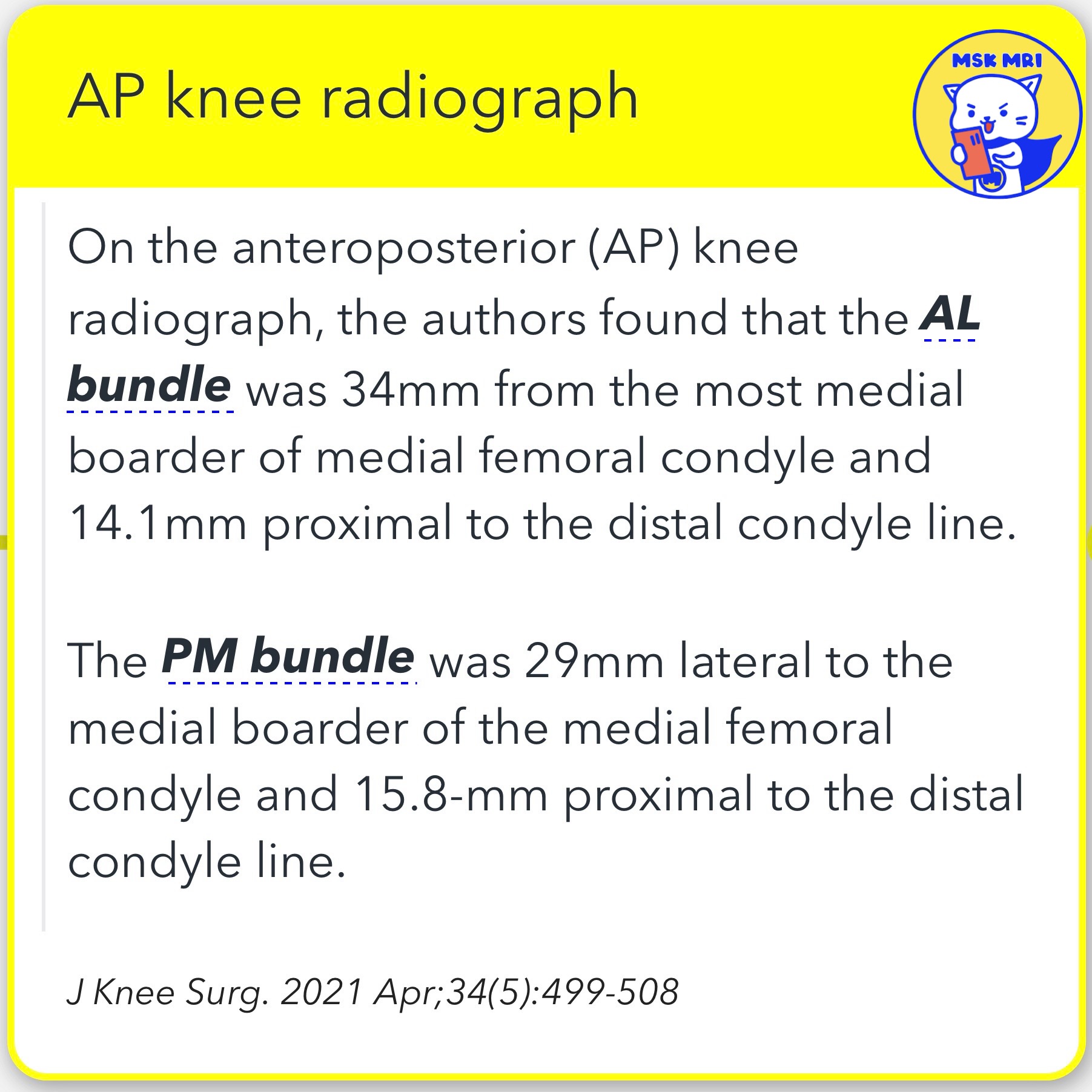
4️⃣ AP Knee Radiograph Findings
- AL Bundle: Positioned 34mm from the medial border of the medial femoral condyle and 14.1mm proximal to the distal condyle line.
- PM Bundle: Located 29mm lateral to the medial border and 15.8mm proximal to the distal condyle line.
- Source: J Knee Surg. 2021 Apr;34(5):499-508.
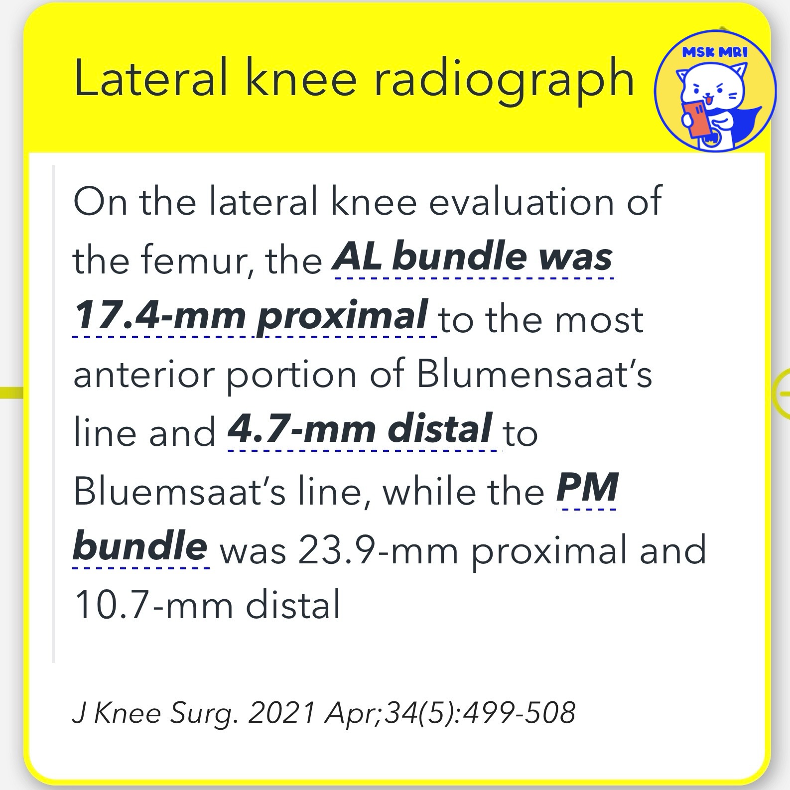
5️⃣ Lateral Knee Radiograph Evaluation
- AL Bundle: Found to be 17.4mm proximal to the most anterior portion of Blumensaat’s line and 4.7mm distal.
- PM Bundle: Positioned 23.9mm proximal and 10.7mm distal to Blumensaat’s line.
- Source: J Knee Surg. 2021 Apr;34(5):499-508.
"Visualizing MSK Radiology: A Practical Guide to Radiology Mastery"
© 2022 MSK MRI Jee Eun Lee All Rights Reserved.
#VisualizingMSK #KneeMRI #PCL #KneeAnatomy
You should not distribute or commercially exploit the content.
You should not transmit or store it on any other website or electronic retrieval system.