Click the link to purchase on Amazon 🎉📚
==============================================
🎥 Check Out All Videos at Once! 📺
👉 Visit Visualizing MSK Blog to explore a wide range of videos! 🩻
https://visualizingmsk.blogspot.com/?view=magazine
📚 You can also find them on MSK MRI Blog and Naver Blog! 📖
https://www.instagram.com/msk_mri/
Click now to stay updated with the latest content! 🔍✨
==============================================
🔴 Meniscofemoral Ligaments 🔴
1️⃣ Femoral Bony Landmarks
- The medial intercondylar ridge marks the superior border of the AL and PM bundle origins.
- The medial bifurcate ridge separates the two bundles.
(J Knee Surg. 2021 Apr;34(5):499-508)
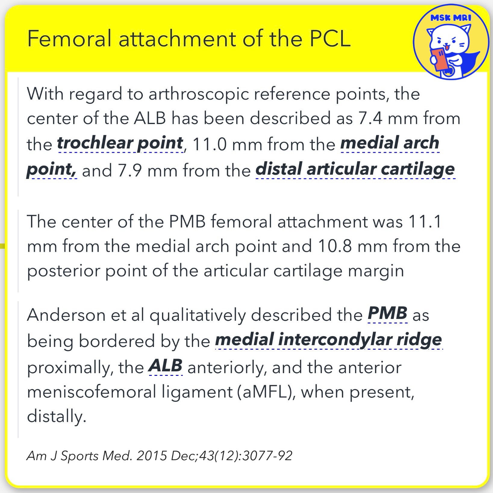
2️⃣ Femoral Attachment of the PCL
- The ALB's center is located 7.4 mm from the trochlear point, 11.0 mm from the medial arch point, and 7.9 mm from the distal articular cartilage.
- The PMB femoral attachment center is 11.1 mm from the medial arch point and 10.8 mm from the posterior point of the articular cartilage margin.
- The PMB is bordered by the medial intercondylar ridge proximally, the ALB anteriorly, and the aMFL distally when present.
(Am J Sports Med. 2015 Dec;43(12):3077-92)
3️⃣ PCL Complex
- The PCL is functionally supported by the anterior (Humphrey) and posterior (Wrisberg) meniscofemoral ligaments and the posterolateral corner structures.
- The function of the MFLs is unclear but hypothesized to act as a secondary restraint, forming the PCL complex with the PCL.
(Magn Reson Imaging Clin N Am. 2022 May;30(2):261-275; Semin Musculoskelet Radiol. 2016 Feb;20(1):43-51)
4️⃣ Meniscofemoral Ligaments (MFLs)
- The anterior and posterior MFL can be observed in 20–75% and 70–100% of knees, respectively, with at least one MFL present in more than 90% of cases.
- On the femoral side, the MFLs attach just proximal (posterior MFL) and distal (anterior MFL) to the PMB's femoral attachment, exhibiting a circular cross-section.
(Knee Surg Sports Traumatol Arthrosc. 2021 Mar;29(3):672-681)
5️⃣ Meniscofemoral Ligaments Details
- The aMFL femoral attachment varies, with 80% of specimens attaching distally to the PMB and the remaining 20% to the ALB.
- The pMFL has a femoral attachment area of 31 mm², located proximal to the medial intercondylar ridge and PMB, and attaches distally to the posterior horn of the lateral meniscus.
(Am J Sports Med. 2015 Dec;43(12):3077-92)
"Visualizing MSK Radiology: A Practical Guide to Radiology Mastery"
© 2022 MSK MRI Jee Eun Lee All Rights Reserved.
#VisualizingMSK #KneeMRI #PCL #KneeAnatomy #PCLanatomy
You should not distribute or commercially exploit the content.
You should not transmit or store it on any other website or electronic retrieval system.