Click the link to purchase on Amazon 🎉📚
==============================================
🎥 Check Out All Videos at Once! 📺
👉 Visit Visualizing MSK Blog to explore a wide range of videos! 🩻
https://visualizingmsk.blogspot.com/?view=magazine
📚 You can also find them on MSK MRI Blog and Naver Blog! 📖
https://www.instagram.com/msk_mri/
Click now to stay updated with the latest content! 🔍✨
==============================================
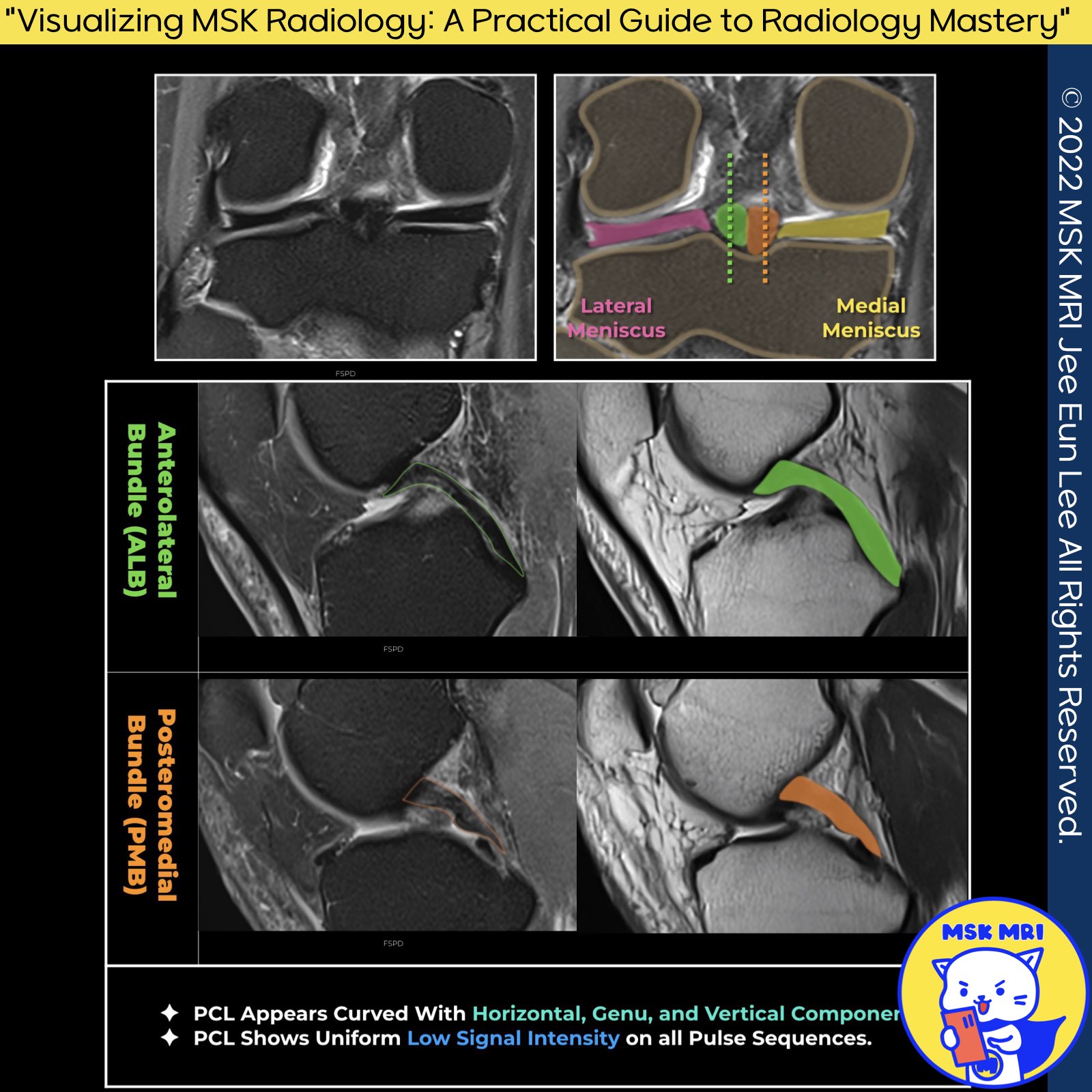
🔴 Anatomy of the PCL on Sagittal Images 🔴
1️⃣ Sagittal Plane Overview
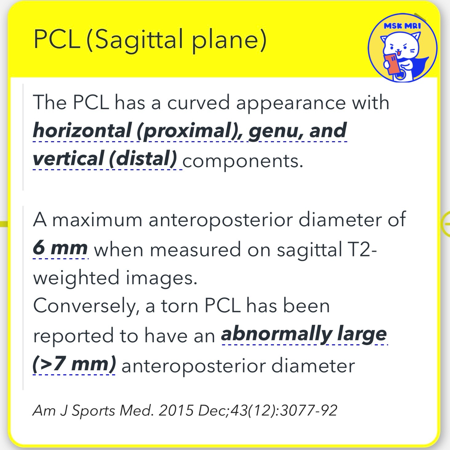
➡️ The Posterior Cruciate Ligament in the sagittal plane displays a curved form, consisting of horizontal proximal, genu, and vertical distal components.
Measurement and Appearance on Sagittal T2-Weighted Images
- Normal PCL Diameter: The PCL shows a maximum anteroposterior diameter of 6 mm on sagittal T2-weighted images.
- Torn PCL Indicators: A PCL tear is indicated by an abnormally large anteroposterior diameter, greater than 7 mm.
Source: American Journal of Sports Medicine, Dec 2015, 43:3077-92
2️⃣ Anterolateral Bundle Characteristics
➡️ The AL bundle, larger at the midsubstance, is distinguished by the positioning of its femoral insertions. The stronger ALB originates anteriorly from the lateral margin of the medial femoral condyle, ahead of the PMB's origin.
Source: Magnetic Resonance Imaging Clinics of North America, Nov 2014, 22:557-80
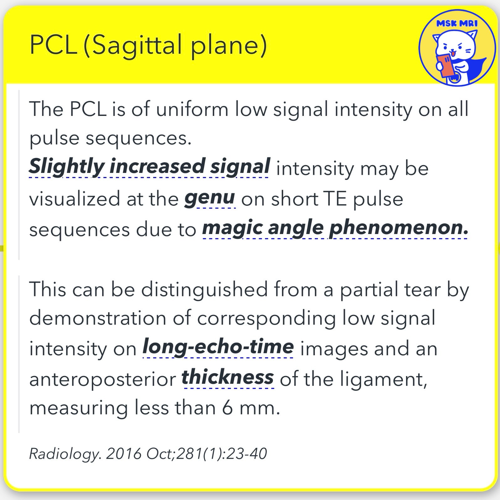
➡️ Uniformity and Signal Intensity
- Uniform Low Signal Intensity: The PCL exhibits uniform low signal intensity across all pulse sequences.
- Increased Signal at the Genu: Slight increase in signal intensity may occur at the genu on short TE pulse sequences, attributed to the magic angle phenomenon.
- Differentiation from Partial Tear: A partial tear can be distinguished by the presence of corresponding low signal intensity on long-echo-time images and an anteroposterior thickness of the ligament measuring less than 6 mm.
Source: Radiology, Oct 2016, 281:23-40
"Visualizing MSK Radiology: A Practical Guide to Radiology Mastery"
© 2022 MSK MRI Jee Eun Lee All Rights Reserved.
#VisualizingMSK #KneeMRI #PCL #KneeAnatomy #PCLanatomy #Humphrey #Wrisberg
You should not distribute or commercially exploit the content.
You should not transmit or store it on any other website or electronic retrieval system.